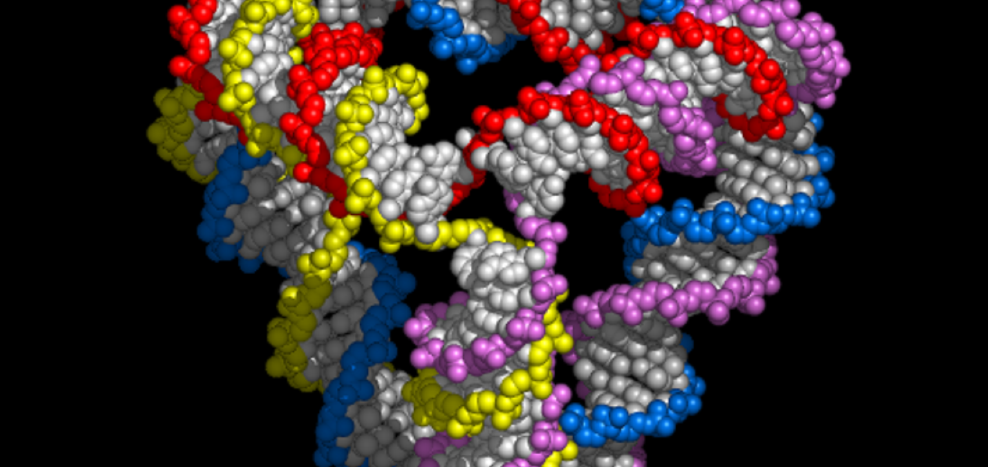
Dimensions and Global Twist of Single-Layer DNA Origami Measured by Small-Angle X-Ray Scattering.
ACS nano (2018)
Abstract:
The rational design of complementary DNA sequences can be used to create nanostructures that self-assemble with nanometer precision. DNA nanostructures have been imaged by atomic force microscopy and electron microscopy. Small-angle X-ray scattering (SAXS) provides complementary structural information on the ensemble-averaged state of DNA nanostructures in solution. Here we demonstrate that SAXS can distinguish between different single-layer DNA origami tiles that look identical when immobilized on a mica surface and imaged with atomic force microscopy. We use SAXS to quantify the magnitude of global twist of DNA origami tiles with different crossover periodicities: these measurements highlight the extreme structural sensitivity of single-layer origami to the location of strand crossovers. We also use SAXS to quantify the distance between pairs of gold nanoparticles tethered to specific locations on a DNA origami tile and use this method to measure the overall dimensions and geometry of the DNA nanostructure in solution. Finally, we use indirect Fourier methods, which have long been used for the interpretation of SAXS data from biomolecules, to measure the distance between DNA helix pairs in a DNA origami nanotube. Together, these results provide important methodological advances in the use of SAXS to analyze DNA nanostructures in solution and insights into the structures of single-layer DNA origami.Chiral DNA origami nanotubes with well‐defined and addressable inside and outside surfaces
Angewandte Chemie International Edition Wiley‐VCH Verlag 57:26 (2018) 7687-7690
Abstract:
We report the design and assembly of chiral DNA nanotubes with well‐defined and addressable inside and outside surfaces. We demonstrate that the outside surface can be functionalised with a chiral arrangement of gold nanoparticles to create a plasmonic device and that the inside surface can be functionalised with a track for a molecular motor allowing transport of a cargo within the central cavity.Self-propulsion of catalytic nanomotors synthesised by seeded growth of asymmetric platinum–gold nanoparticles
Chemical Communications Royal Society of Chemistry 54:15 (2018) 1901-1904
Abstract:
Asymmetric bimetallic nanomotors are synthesised by seeded growth in solution, providing a convenient and high-throughput alternative to the usual top-down lithographic fabrication of self-propelled catalytic nanoparticles. These synthetic nanomotors catalyse H2O2 decomposition and exhibit enhanced diffusion that depends on fuel concentration, consistent with their chemical propulsion.Lipid Bilayer Modulation using DNA Origami Mimics of Clathrin
Biophysical Journal Elsevier 114:3 (2018) 103a
DNA origami nanostructured surfaces for enhanced detection of molecular interactions
22nd International Conference on Miniaturized Systems for Chemistry and Life Sciences, MicroTAS 2018 1 (2018) 16-19