Home
Department Of Physics text logo
  • Research
    • Our research
    • Our research groups
    • Our research in action
    • Research funding support
    • Summer internships for undergraduates
  • Study
    • Undergraduates
    • Postgraduates
  • Engage
    • For young people
    • For teachers
    • For the public
    • For alumni
    • For business
  • Support
Menu
CMP
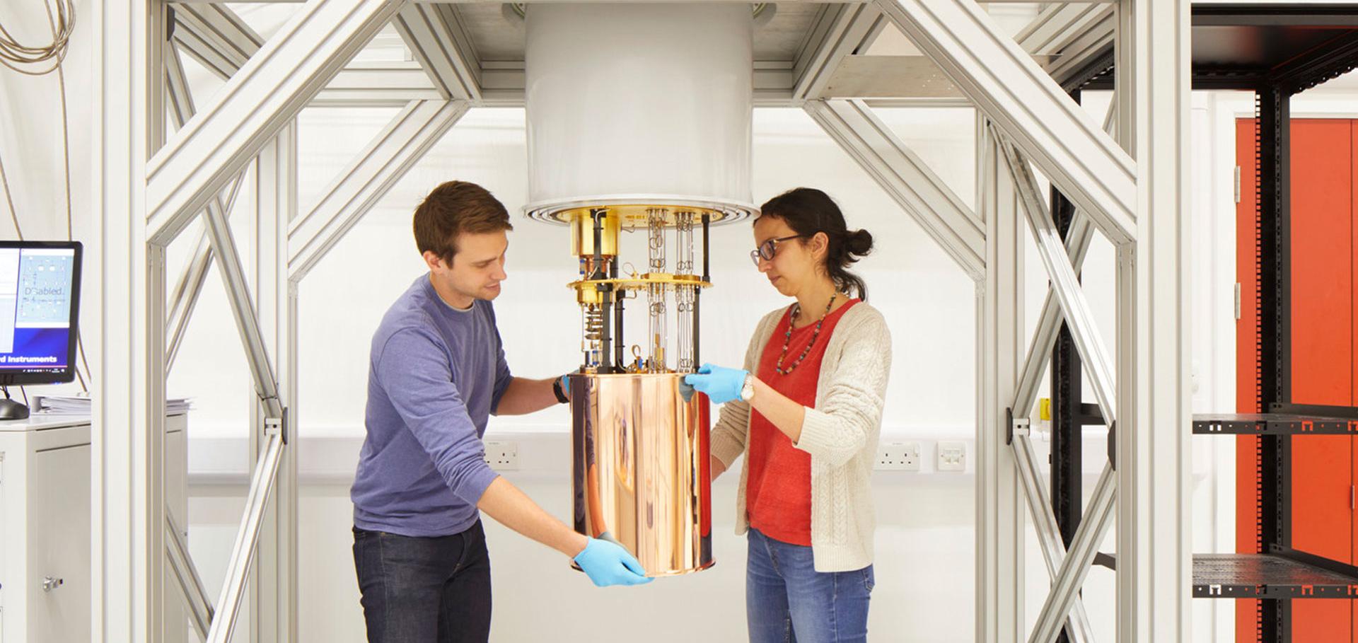
Credit: Jack Hobhouse

Anuj Tiwari

PDRA in Single-Cell Imaging of Antimicrobial Resistance

Sub department

  • Condensed Matter Physics
anuj.tiwari@physics.ox.ac.uk
  • About
  • Publications

Droplet-based methodology for investigating bacterial population dynamics in response to phage exposure

Frontiers in Microbiology Frontiers Media 14 (2023) 1260196-1260196

Authors:

Nela Nikolic, Vasileios Anagnostidis, Anuj Tiwari, Remy Chait, Fabrice Gielen

Abstract:

An alarming rise in antimicrobial resistance worldwide has spurred efforts into the search for alternatives to antibiotic treatments. The use of bacteriophages, bacterial viruses harmless to humans, represents a promising approach with potential to treat bacterial infections (phage therapy). Recent advances in microscopy-based single-cell techniques have allowed researchers to develop new quantitative methodologies for assessing the interactions between bacteria and phages, especially the ability of phages to eradicate bacterial pathogen populations and to modulate growth of both commensal and pathogen populations. Here we combine droplet microfluidics with fluorescence time-lapse microscopy to characterize the growth and lysis dynamics of the bacterium Escherichia coli confined in droplets when challenged with phage. We investigated phages that promote lysis of infected E. coli cells, specifically, a phage species with DNA genome, T7 (Escherichia virus T7) and two phage species with RNA genomes, MS2 (Emesvirus zinderi) and Qβ (Qubevirus durum). Our microfluidic trapping device generated and immobilized picoliter-sized droplets, enabling stable imaging of bacterial growth and lysis in a temperature-controlled setup. Temporal information on bacterial population size was recorded for up to 25 h, allowing us to determine growth rates of bacterial populations and helping us uncover the extent and speed of phage infection. In the long-term, the development of novel microfluidic single-cell and population-level approaches will expedite research towards fundamental understanding of the genetic and molecular basis of rapid phage-induced lysis and eco-evolutionary aspects of bacteria-phage dynamics, and ultimately help identify key factors influencing the success of phage therapy
More details from the publisher
Details from ORA
More details
More details

SC-MAPS: A Multichannel Single-Cell Microscopy Dataset for Deep Learning-Based Bacterial Species Identification and Antimicrobial Susceptibility Testing Routing

Authors:

ANUJ TIWARI, Piers Turner

Abstract:

This repository contains the SC-MAPS (Single-Cell Microscopy for Antimicrobial Prediction and Species identification) dataset, a large-scale, multichannel bacterial microscopy resource developed to support deep learning-based bacterial species identification, phenotypic characterisation and classification into antimicrobial susceptibility testing (AST)-routing groups. The dataset is provided as two separate ZIP archives: 1. SC-MAPS Primary Dataset (13 bacterial species) The primary dataset comprises 916,435 segmented single bacterial cells representing 13 clinically relevant bacterial species, collected across 49 species–strain biological replicates. The dataset includes multiple independent strains per species, enabling evaluation of model generalisation to previously unseen strains. Individual bacterial cells were segmented from microscopy images and extracted as standardised 96 × 96-pixel image crops while preserving their native imaging scale. Images were acquired using complementary bright-field, phase-contrast and fluorescence microscopy channels, including DAPI (nucleic acids), WGA-AF647 (cell-envelope-associated structures) and FM1-43 (membrane staining). Multichannel images are stored as NumPy (.npy) arrays for computational analysis. The dataset was used to develop and evaluate convolutional neural network classifiers for bacterial species identification and classification into eight clinically informed AST-routing groups. Training and validation were performed using development strains, while independent strains were reserved for holdout evaluation. 2. SC-MAPS Additional Species Dataset (5 previously unseen species) The second archive contains single-cell microscopy data from five additional bacterial species excluded from the original 13-species model training dataset. These images were processed using the SC-MAPS single-cell extraction workflow and provide an independent resource for investigating model behaviour when encountering organisms outside the training classes. The dataset supports investigations of learned phenotypic representations, cross-species relationships, classification uncertainty and out-of-distribution detection. Dataset Applications Together, these archives provide a resource for developing and benchmarking computational approaches to bacterial identification, multichannel image classification, phenotypic feature extraction, strain-level generalisation, population-level prediction aggregation and open-set recognition. The accompanying SC-MAPS analysis and model-training code is maintained separately on GitHub to facilitate reproducibility and further development of microscopy-based bacterial classification approaches.

Pagination

  • First page First
  • Previous page Prev
  • Page 1
  • Current page 2

Footer Menu

  • Contact us
  • Giving to the Dept of Physics
  • Work with us
  • Media

User account menu

  • Log in

Follow us

FIND US

Clarendon Laboratory,

Parks Road,

Oxford,

OX1 3PU

CONTACT US

Tel: +44(0)1865272200

University of Oxfrod logo Department Of Physics text logo
IOP Juno Champion logo Athena Swan Silver Award logo

© University of Oxford - Department of Physics

Cookies | Privacy policy | Accessibility statement

Built by: Versantus

  • Home
  • Research
  • Study
  • Engage
  • Our people
  • News & Comment
  • Events
  • Our facilities & services
  • About us
  • Giving to Physics
  • Current students
  • Staff intranet