
VHEE FLASH sparing effect measured at CLEAR, CERN with DNA damage of pBR322 plasmid as a biological endpoint
Scientific Reports Nature Research 14:1 (2024) 14803
Abstract:
Ultra-high dose rate (UHDR) irradiation has been shown to have a sparing effect on healthy tissue, an effect known as ‘FLASH’. This effect has been studied across several radiation modalities, including photons, protons and clinical energy electrons, however, very little data is available for the effect of FLASH with Very High Energy Electrons (VHEE). pBR322 plasmid DNA was used as a biological model to measure DNA damage in response to Very High Energy Electron (VHEE) irradiation at conventional (0.08 Gy/s), intermediate (96 Gy/s) and ultra-high dose rates (UHDR, (2 × 10⁹ Gy/s) at the CERN Linear Electron Accelerator (CLEAR) user facility. UHDRs were used to determine if the biological FLASH effect could be measured in the plasmid model, within a hydroxyl scavenging environment. Two different concentrations of the hydroxyl radical scavenger Tris were used in the plasmid environment to alter the proportions of indirect damage, and to replicate a cellular scavenging capacity. Indirect damage refers to the interaction of ionising radiation with molecules and species to generate reactive species which can then attack DNA. UHDR irradiated plasmid was shown to have significantly reduced amounts of damage in comparison to conventionally irradiated, where single strand breaks (SSBs) was used as the biological endpoint. This was the case for both hydroxyl scavenging capacities. A reduced electron energy within the VHEE range was also determined to increase the DNA damage to pBR322 plasmid. Results indicate that the pBR322 plasmid model can be successfully used to explore and test the effect of UHDR regimes on DNA damage. This is the first study to report FLASH sparing with VHEE, with induced damage to pBR322 plasmid DNA as the biological endpoint. UHDR irradiated plasmid had reduced amounts of DNA single-strand breaks (SSBs) in comparison with conventional dose rates. The magnitude of the FLASH sparing was a 27% reduction in SSB frequency in a 10 mM Tris environment and a 16% reduction in a 100 mM Tris environment.CERN-based experiments and Monte-Carlo studies on focused dose delivery with very high energy electron (VHEE) beams for radiotherapy applications
Scientific Reports Nature Research 14:1 (2024) 11120
Abstract:
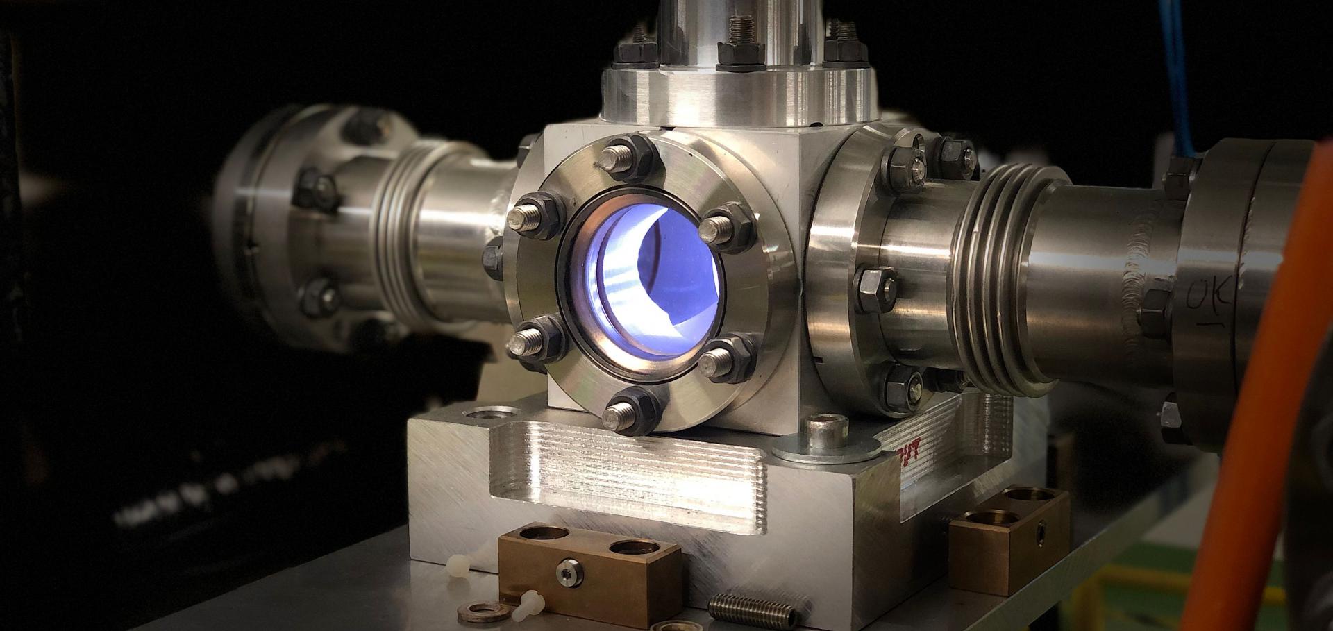
Very High Energy Electron (VHEE) beams are a promising alternative to conventional radiotherapy due to their highly penetrating nature and their applicability as a modality for FLASH (ultra-high dose-rate) radiotherapy. The dose distributions due to VHEE need to be optimised; one option is through the use of quadrupole magnets to focus the beam, reducing the dose to healthy tissue and allowing for targeted dose delivery at conventional or FLASH dose-rates. This paper presents an in depth exploration of the focusing achievable at the current CLEAR (CERN Linear Electron Accelerator for Research) facility, for beam energies >200 MeV. A shorter, more optimal quadrupole setup was also investigated using the TOPAS code in Monte Carlo simulations, with dimensions and beam parameters more appropriate to a clinical situation. This work provides insight into how a focused VHEE radiotherapy beam delivery system might be achieved.Development of a novel fibre optic beam profile and dose monitor for very high energy electron radiotherapy at ultrahigh dose rates
Physics in Medicine & Biology IOP Publishing 69:8 (2024) 085006-085006
Abstract:
Very High Energy Electrons (VHEE) are a promising radiotherapy modality due to their increased penetration, reduced sensitivity to inhomogeneities, and delivery via scanning or focusing. VHEE beams at ultrahigh dose rates (UHDR) could be beneficial for treating deep-seated tumours using the FLASH effect, which selectively spares healthy tissues while maintaining effective tumour control. One of the main challenges in making VHEE FLASH treatment clinically viable is real-time dosimetry and beam monitoring, as ionisation chambers exhibit non-linear responses at UHDR due to recombination effects. This research addresses this challenge through the characterisation of VHEE interactions using Monte Carlo (MC) simulations, film dosimetry at the CLEAR Facility, and by developing a novel fibre array beam monitor. Using TOPAS MC simulations, the interactions of VHEE beams were characterised. The dose distributions and resulting secondary particles generated from these interactions were evaluated to determine feasible in vivo dose verification methods for VHEE UHDR beams. A radiochromic film dosimetry protocol was developed for VHEE FLASH experiments at the CLEAR Facility to ensure accurate dose measurements. Various Gaussian beam size determination methods were compared. Charge measurements using an integrated current transformer were correlated with dose-area-product measurements on radiochromic films for both UHDR and conventional irradiations. Radiochromic film measurements were also compared to those made with other passive dosimeters to ensure accuracy and reliability. A novel optical fibre beam monitor was developed for real-time beam profile and dose monitoring at UHDR with VHEE beams. Consisting of silica fibres and a CMOS camera, the monitor was tested and characterised at the CLEAR Facility. A linear response with dose rate was demonstrated alongside accurate beam profile measurements for Gaussian and uniform beams. This shows real potential as a solution to address the critical need for accurate beam monitoring with VHEE FLASH radiotherapyMini-GRID radiotherapy on the CLEAR very-high-energy electron beamline: collimator optimization, film dosimetry, and Monte Carlo simulations
Physics in Medicine and Biology IOP Publishing 69:5 (2024) 055003
Plastic Scintillator Dosimetry of Ultrahigh Dose-Rate 200 MeV Electrons at CLEAR
IEEE Sensors Journal Institute of Electrical and Electronics Engineers (IEEE) 24:9 (2024) 14229-14237