Home
Department Of Physics text logo
  • Research
    • Our research
    • Our research groups
    • Our research in action
    • Research funding support
    • Summer internships for undergraduates
  • Study
    • Undergraduates
    • Postgraduates
  • Engage
    • For alumni
    • For business
    • For schools
    • For the public
Menu
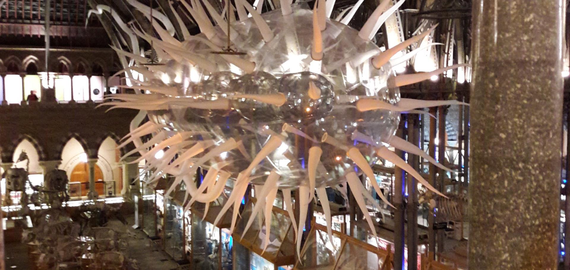
Giant Inflatable. E coli model

Inflatable model of E. coli installed at the Natural History Museum, Oxford, as part of the the 'Bacterial World' exhibition

Helen Miller

Long Term Visitor

Sub department

  • Condensed Matter Physics
helen.miller@physics.ox.ac.uk
Orcid profile
Google scholar
  • About
  • Publications

An automated image analysis framework for segmentation and division plane detection of single live Staphylococcus aureus cells which can operate at millisecond sampling time scales using bespoke Slimfield microscopy

Physical Biology 13:5 (2016) 055002-055002

Authors:

AJM Wollman, H Miller, S Foster, MC Leake
More details from the publisher
Details from ORA
More details
More details

Developing a single-molecule fluorescence tool to quantify DNA damage

Biophysical Journal Elsevier 110:3 suppl. 1 (2016) p164a

Abstract:

Quantification of DNA damage is an important technique for medical physics, for example to assess damage caused by the quinolone antibiotics, or to examine the effects of novel cancer treatments such as low-temperature plasma therapy on healthy or tumorous cells. Existing damage quantification techniques such as the alkaline comet assay [1] are often subjective in their results, especially at higher damage levels. Methods using immunofluorescence [2] often lose information due to the three dimensional nature of the cell.
More details from the publisher
Details from ORA
More details

Designing a Single-Molecule Biophysics Tool for Characterising DNA Damage for Techniques that Kill Infectious Pathogens Through DNA Damage Effects.

Advances in experimental medicine and biology 915 (2016) 115-127

Authors:

Helen Miller, Adam JM Wollman, Mark C Leake

Abstract:

Antibiotics such as the quinolones and fluoroquinolones kill bacterial pathogens ultimately through DNA damage. They target the essential type IIA topoisomerases in bacteria by stabilising the normally transient double-strand break state which is created to modify the supercoiling state of the DNA. Here we discuss the development of these antibiotics and their method of action. Existing methods for DNA damage visualisation, such as the comet assay and immunofluorescence imaging can often only be analysed qualitatively and this analysis is subjective. We describe a putative single-molecule fluorescence technique for quantifying DNA damage via the total fluorescence intensity of a DNA origami tile fully saturated with an intercalating dye, along with the optical requirements for how to implement these into a light microscopy imaging system capable of single-molecule millisecond timescale imaging. This system promises significant improvements in reproducibility of the quantification of DNA damage over traditional techniques.
More details from the publisher
More details
More details

Superresolution imaging of single DNA molecules using stochastic photoblinking of minor groove and intercalating dyes.

Methods (San Diego, Calif.) 88 (2015) 81-88

Authors:

Helen Miller, Zhaokun Zhou, Adam JM Wollman, Mark C Leake

Abstract:

As proof-of-principle for generating superresolution structural information from DNA we applied a method of localization microscopy utilizing photoblinking comparing intercalating dye YOYO-1 against minor groove binding dye SYTO-13, using a bespoke multicolor single-molecule fluorescence microscope. We used a full-length ∼49 kbp λ DNA construct possessing oligo inserts at either terminus allowing conjugation of digoxigenin and biotin at opposite ends for tethering to a glass coverslip surface and paramagnetic microsphere respectively. We observed stochastic DNA-bound dye photoactivity consistent with dye photoblinking as opposed to binding/unbinding events, evidenced through both discrete simulations and continuum kinetics analysis. We analyzed dye photoblinking images of immobilized DNA molecules using superresolution reconstruction software from two existing packages, rainSTORM and QuickPALM, and compared the results against our own novel home-written software called ADEMS code. ADEMS code generated lateral localization precision values of 30-40 nm and 60-70 nm for YOYO-1 and SYTO-13 respectively at video-rate sampling, similar to rainSTORM, running more slowly than rainSTORM and QuickPALM algorithms but having a complementary capability over both in generating automated centroid distribution and cluster analyses. Our imaging system allows us to observe dynamic topological changes to single molecules of DNA in real-time, such as rapid molecular snapping events. This will facilitate visualization of fluorescently-labeled DNA molecules conjugated to a magnetic bead in future experiments involving newly developed magneto-optical tweezers combined with superresolution microscopy.
More details from the publisher
More details
More details

Developing a New Biophysical Tool to Combine Magneto-Optical Tweezers with Super-Resolution Fluorescence Microscopy

Photonics MDPI 2:3 (2015) 758-772

Authors:

Zhaokun Zhou, Helen Miller, Adam Wollman, Mark Leake
More details from the publisher

Pagination

  • First page First
  • Previous page Prev
  • Page 1
  • Page 2
  • Current page 3
  • Page 4
  • Next page Next
  • Last page Last

Footer Menu

  • Contact us
  • Giving to the Dept of Physics
  • Work with us
  • Media

User account menu

  • Log in

Follow us

FIND US

Clarendon Laboratory,

Parks Road,

Oxford,

OX1 3PU

CONTACT US

Tel: +44(0)1865272200

University of Oxfrod logo Department Of Physics text logo
IOP Juno Champion logo Athena Swan Silver Award logo

© University of Oxford - Department of Physics

Cookies | Privacy policy | Accessibility statement

Built by: Versantus

  • Home
  • Research
  • Study
  • Engage
  • Our people
  • News & Comment
  • Events
  • Our facilities & services
  • About us
  • Current students
  • Staff intranet