Home
Department Of Physics text logo
  • Research
    • Our research
    • Our research groups
    • Our research in action
    • Research funding support
    • Summer internships for undergraduates
  • Study
    • Undergraduates
    • Postgraduates
  • Engage
    • For alumni
    • For business
    • For schools
    • For the public
Menu
CMP
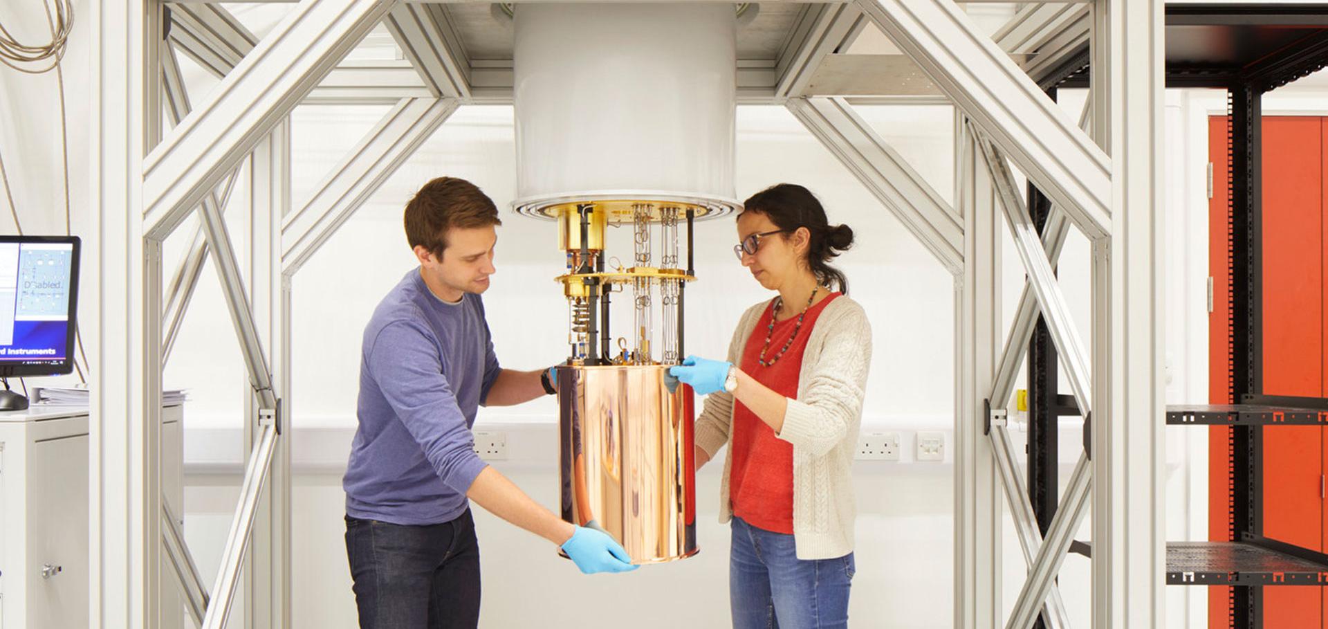
Credit: Jack Hobhouse

Peter Proks

Postdoctoral Research Assistant

Sub department

  • Condensed Matter Physics
peter.proks@physics.ox.ac.uk
Telephone: 72426
Clarendon Laboratory, room 071.4 & 071.7
  • About
  • Publications

The role of the C-terminus of SUR in the differential regulation of beta-cell and cardiac KATP channels by MgADP and metabolism

Journal of Physiology John Wiley & Sons, Inc 596:24 (2018) 6205-6217

Authors:

Natascia Vedovato, Olof Rorsman, Konstantin Hennis, Frances Ashcroft, Peter Proks

Abstract:

ATP‐sensitive potassium (KATP) channels couple the metabolic state of a cell to its electrical activity and play important physiological roles in many tissues. In contrast to β‐cell (Kir6.2/SUR1) channels, which open when extracellular glucose levels fall, cardiac (Kir6.2/SUR2A) channels remain closed. It is known this is due to differences in the SUR subunit rather than cell metabolism. As ATP inhibition and MgADP activation are similar for both types of channels, we investigated channel inhibition by MgATP in the presence of 100 μm MgADP immediately after patch excision (when the channel open probability (PO) is near maximal). The results were strikingly different: 100 μm MgADP substantially reduced MgATP inhibition of Kir6.2/SUR1, but had no effect on MgATP inhibition of Kir6.2/SUR2A. Exchanging the final 42 residues of SUR2A with that of SUR1 switched the channel phenotype (and v.v.), and deleting this region abolished Mg‐nucleotide activation. This suggests the C‐terminal 42 residues are important for the ability of MgADP to influence ATP inhibition at Kir6.2. This region was also necessary, although, not sufficient for activation of the KATP channel in the intact cells by metabolic inhibition (azide). We conclude that the ability of MgADP to impair ATP inhibition at Kir6.2 accounts, in part, for the differential metabolic sensitivities of β‐cell and cardiac KATP channels.
More details from the publisher
Details from ORA
More details
More details

Binding of sulphonylureas to plasma proteins – a KATP channel perspective

PLoS ONE Public Library of Science 13:5 (2018) e0197634

Authors:

Peter Proks, Holger Kramer, Elizabeth Haythorne, Frances Ashcroft

Abstract:

Sulphonylurea drugs stimulate insulin secretion from pancreatic β-cells primarily by inhibiting ATP sensitive potassium (KATP) channels in the β-cell membrane. The effective sulphonylurea concentration at its site of action is significantly attenuated by binding to serum albumin, which makes it difficult to compare in vitro and in vivo data. We therefore measured the ability of gliclazide and glibenclamide to inhibit KATP channels and stimulate insulin secretion in the presence of serum albumin. We used this data, together with estimates of free drug concentrations from binding studies, to predict the extent of sulphonylurea inhibition of KATP channels at therapeutic concentrations in vivo. KATP currents from mouse pancreatic β-cells and Xenopus oocytes were measured using the patch-clamp technique. Gliclazide and glibenclamide binding to human plasma were determined in spiked plasma samples using an ultrafiltration-mass spectrometry approach. Bovine serum albumin (60g/l) produced a mild, non-significant reduction of gliclazide block of KATP currents in pancreatic β-cells and Xenopus oocytes. In contrast, glibenclamide inhibition of recombinant KATP channels was dramatically suppressed by albumin (predicted free drug concentration <0.1%). Insulin secretion was also reduced. Free concentrations of gliclazide and glibenclamide in the presence of human plasma measured in binding experiments were 15% and 0.05%, respectively. Our data suggest the free concentration of glibenclamide in plasma is too low to account for the drug’s therapeutic effect. In contrast, the free gliclazide concentration in plasma is high enough to close KATP channels and stimulate insulin secretion.
More details from the publisher
Details from ORA
More details
More details

Differential metabolic and nucleotide sensitivity of beta-cell and cardiac K-ATP channels

Biophysical Journal Cell Press 114:3, S1 (2018) 203a-204a

Authors:

Natascia Vedovato, Peter Proks, Olof H Rorsman, K Hennis, Frances M Ashcroft
More details from the publisher
Details from ORA
More details
More details

Correction to 'Running out of time: the decline of channel activity and nucleotide activation in adenosine triphosphate-sensitive K-channels'.

Philosophical Transactions B: Biological Sciences Royal Society 371:1702 (2016) 20160330

Authors:

Peter Proks, Michael Puljung, Natascia Vedovato, Gregor Sachse, Rachel Mulvaney, Frances Ashcroft
More details from the publisher
Details from ORA
More details
More details

Running out of time: the decline of channel activity and nucleotide activation in ATP-sensitive K-channels

Philosophical Transactions of the Royal Society of London: Biological Sciences Royal Society 371:1700 (2016) 20150426

Authors:

Peter Proks, Michael C Puljung, Natascia Vedovato, Gregor Sachse, Rachel Mulvaney, Frances Ashcroft

Abstract:

KATP channels act as key regulators of electrical excitability by coupling metabolic cues - mainly intracellular adenine nucleotide concentrations - to cellular potassium ion efflux. However, their study has been hindered by their rapid loss of activity in excised membrane patches (rundown), and by a second phenomenon, the decline of activation by Mg-nucleotides (DAMN). Degradation of PI(4,5)P2 and other phosphoinositides is the strongest candidate for the molecular cause of rundown. Broad evidence indicates that most other determinants of rundown (e.g. de-/phosphorylation, intracellular calcium, channel mutations that affect rundown) also act by influencing KATP channel regulation by phosphoinositides. Unfortunately, experimental conditions that reproducibly prevent rundown have remained elusive, necessitating post-hoc data compensation. Rundown is clearly distinct from DAMN. While the former is associated with pore-forming Kir6.2 subunits, DAMN is generally a slower process involving the regulatory sulfonylurea receptor (SUR) subunits. We speculate it arises when SUR subunits enter non-physiological conformational states associated with the loss of SUR nucleotide-binding domain dimerization following prolonged exposure to nucleotide-free conditions. This review presents new information on both rundown and DAMN, summarizes our current understanding of these processes and considers their physiological roles.
More details from the publisher
Details from ORA
More details
More details

Pagination

  • First page First
  • Previous page Prev
  • Page 1
  • Page 2
  • Page 3
  • Current page 4
  • Page 5
  • Page 6
  • Page 7
  • Page 8
  • Page 9
  • …
  • Next page Next
  • Last page Last

Footer Menu

  • Contact us
  • Giving to the Dept of Physics
  • Work with us
  • Media

User account menu

  • Log in

Follow us

FIND US

Clarendon Laboratory,

Parks Road,

Oxford,

OX1 3PU

CONTACT US

Tel: +44(0)1865272200

University of Oxfrod logo Department Of Physics text logo
IOP Juno Champion logo Athena Swan Silver Award logo

© University of Oxford - Department of Physics

Cookies | Privacy policy | Accessibility statement

Built by: Versantus

  • Home
  • Research
  • Study
  • Engage
  • Our people
  • News & Comment
  • Events
  • Our facilities & services
  • About us
  • Current students
  • Staff intranet