Achieving flexible competence: bridging the investment dichotomy between infectious diseases and cancer
BMJ Global Health BMJ 5:12 (2020) e003252
Arbitrarily large tomography with iterative algorithms on multiple GPUs using the TIGRE toolbox
Journal of Parallel and Distributed Computing Elsevier 146 (2020) 52-63
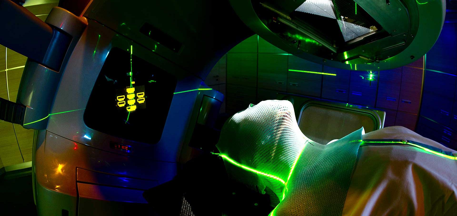
Overcoming Challenges in Providing Radiation Therapy to Patients With Cancer in Nigeria and Experience in the National Hospital Abuja, Nigeria
JCO Global Oncology American Society of Clinical Oncology (ASCO) 6:6 (2020) jgo.19.00177
State – of – the – art and the future of particle therapy (Perspectives for the see countries)
Annals of the University of Craiova, Physics 30:2 (2020) 246-262
Abstract:
Radiation therapy (RT) is aimed to treat cancer cells with a radiation dose sufficient to stop their growth and simultaneously to spare the surrounding healthy tissue. Hadron Therapy (HT) alternatively called Particle Therapy (PT) involves accelerating hadrons (charged particles such as protons or heavier ions) to almost the speed of light, then “painting” the tumour’s volume precisely with the radiation beam. Advances are continuously being made with the developments of new and more accurate technologies. In this work, we intend to show the advancement in the PT and the prospective advancements beyond the-state-of-the-art in (1) accelerator technologies that provide higher intensity ion beams, improvement in the ion-beam “optics” and detection technologies, (2) new gantry design, (3) radiobiology innovative research of the lethal and DNA recovery effects on different accelerated ions species on radioresistant cancer cell lines, and also an examination of possibilities of FLASH PT treatments (using higher doses at a reduced number of treatments), (4) detection and imaging improvements and (5) performances of the profound clinical studies with a big-data approach, in which a comparison between the conventional RT and the PT on large groups of patients that suffer from some types of cancer was made. Cancer is a critical societal issue and currently, it is the second leading cause of death and radiation therapy (RT) is a fundamental component of effective cancer treatment. The main goal of RT is to maximise the damage to the tumour while minimising the damage to the surrounding healthy tissue. The most frequently used modalities of RT use high-energy (MeV) photon or electron beams. Conventional X-ray radiation therapy is characterised by almost exponential attenuation and absorption, and consequently delivers the maximum energy near the beam entrance but continues to deposit significant energy at distances beyond the cancer target. Hadron therapy or Particle therapy (PT) (protons and other light ions) can overcome the limitations of X-rays since hadrons/particles deposit most of their energy at the end of their range and these beams can be shaped with great precision. Hence it allows for a more accurate treatment of the tumour destroying the cancer cells more precisely with minimal damage to surrounding tissue, therefore, sparing the healthy surrounding tissue. The use of protons and carbon-ions for treating cancer has grown over the last 20 years. However, despite the efforts made on compactness and cost reduction, the equipment is still relatively large and expensive, making such facilities economically challenging for most hospitals. To achieve more cost-efficient facilities and thereby give improved access to patients globally, research needs to be strengthened, broadened and combined in several fields including (1) accelerator technologies that provide higher intensity ion beams and improvement in the ion-beam “optics” (2) new gantry design, (3) radiobiological innovative research including the possibilities of using FLASH PT (using high doses in very short irradiation times), (4) detection and imaging improvements and (5) extensive clinical trials with a larger number of patients, in which comparisons between the conventional RT and the PT can be made.Optics Design and Beam Dynamics simulation for a VHEE Radiobiology beam line at PRAE accelerator
Journal of Physics Conference Series IOP Publishing 1350:1 (2019) 012200