Home
Department Of Physics text logo
  • Research
    • Our research
    • Our research groups
    • Our research in action
    • Research funding support
    • Summer internships for undergraduates
  • Study
    • Undergraduates
    • Postgraduates
  • Engage
    • For alumni
    • For business
    • For schools
    • For the public
Menu
website contera

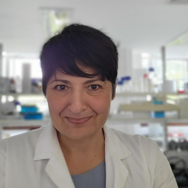
Prof Sonia Antoranz Contera

Professor of Biological Physics

Sub department

  • Condensed Matter Physics
Sonia.AntoranzContera@physics.ox.ac.uk
Telephone: 01865 (2)72269
Clarendon Laboratory, room 208
  • About
  • Publications
Conversation on physics bioinspired materials and the future of architecture
link to video of conversation with architect Amanda Levete on biophysics and the future of architecture

Interactions between transmembrane beta-amyloid(1-40) and phosphatidylcholine bilayers

BIOPHYS J 88:1 (2005) 421A-421A

Authors:

MRR de Planque, GP Mendes, SA Contera, DTS Rijkers, JF Ryan, A Watts
More details

AFM and QCM-D studies in liquid of fibrinogen and vitronectin on Ti and Ta surfaces

Transactions - 7th World Biomaterials Congress (2004) 788

Authors:

AG Hemmersam, SA Contera, M Foss, JK Jensen, P Andreasen, F Besenbacher

Abstract:

The adsorption of fibrinogen followed by specific fibrinogen anti-fibrinogen reaction from a tris-buffered solution at 37°C onto Au, Ta and Ti surfaces was investigated. The concentrations of fibrinogen used were 1 mg/ml, which were close to that found in blood and 0.03 mg/ml. It was found that very small amount of silver on the gold surface have a large effect on the amount of fibrinogen adsorbed. Globular monomeric and polymeric vitronectin proteins for low concentrations were identified using atomic force microscopy (AFM) in liquid.

Effect of time and micropatterns on the behaviour of preosteoblastic cells

Transactions - 7th World Biomaterials Congress (2004) 1363

Authors:

J Justesen, LK Andersen, S Antoranz Contera, M Duch, O Hansen, M Lorentzen, J Chevallier, M Foss, FS Pedersen, F Besenbacher

Abstract:

The influence of the micropatterning of surfaces (in relation to their spreading and morphology) on the behavior of osteoblasts on different surfaces was investigated. Micropatterns were seen to be etched into silicon plates by photolithograhy and coated with a 250 nm tantalum layer. The samples were analyzed by Cryo-SEM (Scanning Electron Microscopy) (CamScan MaXim 2040). The elongation of cells was observed to be faster on deep line patterns than on shallow line patterns.

Role of the Trans-activation Response Element in Dimerization of HIV-1 RNA

Journal of Biological Chemistry 279 (2004) 22243 – 22249

Authors:

S Antoranz Contera, B. Knudsen, C.K.Damgaard, E.S.Andersen
More details from the publisher
More details
More details

Ambient STM and in situ AFM study of nitrite reductase proteins adsorbed on gold and graphite: influence of the substrate on protein interactions.

Ultramicroscopy 97:1-4 (2003) 65-72

Authors:

S Antoranz Contera, H Iwasaki, S Suzuki

Abstract:

Trimeric Achromobacter cycloclastes Cu-containing nitrite reductase (CuNIR) proteins adsorbed on gold and graphite have been studied by ambient STM and in situ AFM. STM resolves them individually and in layers, distinguishing the sub-molecular individual units of the trimer. The Cu atoms are not visible to STM. STM shows that individual CuNIR denatures as it adsorbs on Au, although a deformed trimeric shape can be identified in some cases. CuNIR forms disordered layers on gold. On graphite, ordered self-assembled layers of CuNIR have been resolved by in situ AFM and ambient STM forming parallel rows whose separation distance corresponds to the size of one of the units of the trimer, 5nm. Ambient STM can achieve better resolution than in situ AFM in the images of the layers. We observe differences between domains showing the parallel row structure and unstructured parts of the CuNIR layer by in situ phase imaging AFM.
More details from the publisher
More details

Pagination

  • First page First
  • Previous page Prev
  • …
  • Page 13
  • Page 14
  • Page 15
  • Page 16
  • Page 17
  • Current page 18
  • Page 19
  • Page 20
  • Page 21
  • Next page Next
  • Last page Last

Footer Menu

  • Contact us
  • Giving to the Dept of Physics
  • Work with us
  • Media

User account menu

  • Log in

Follow us

FIND US

Clarendon Laboratory,

Parks Road,

Oxford,

OX1 3PU

CONTACT US

Tel: +44(0)1865272200

University of Oxfrod logo Department Of Physics text logo
IOP Juno Champion logo Athena Swan Silver Award logo

© University of Oxford - Department of Physics

Cookies | Privacy policy | Accessibility statement

Built by: Versantus

  • Home
  • Research
  • Study
  • Engage
  • Our people
  • News & Comment
  • Events
  • Our facilities & services
  • About us
  • Current students
  • Staff intranet