
Using collimator rotation to retain conformal dose distributions with thicker MLC leaves.
Biomedical Physics & Engineering Express IOP Publishing (2026)
Abstract:
The multi-leaf collimator (MLC) is a critical component of modern X-ray radiotherapy systems which allows for the delivery of a conformal dose distribution to the tumour. However, MLCs are one of the most sensitive components in a linac since they contain several components that are prone to failure. The current trend in design is to produce MLCs with ever-increasing numbers of leaves to improve their ability to shape the beam, but this further reduces their robustness. This robustness has shown to be an important factor in low- and middle-income countries (LMICs) where there are currently limited numbers of available linacs per person and that linac downtime has a much greater impact on treatment capacity than in high-income countries. In investigating designs for a more robust MLC system, we assess a new method to deliver a similar conformal tumour dose distribution using a significantly simplified MLC with thicker leaves whilst utilising multiple collimator angles for each delivery to attempt to retain excellent conformity of the dose. The open-source treatment planning software, matRad, was used to generate representative treatment plans to compare the conformity index, homogeneity index and mean dose delivered to circular and crescent-shaped targets for a range of MLC leaf thicknesses and collimator angles. Using MLCs with a leaf thickness of up to 1.5 cm thickness, it was possible to increase the dose conformity by using between 2 to 6 different collimators angles to deliver multiple x-ray beams and generate a conformal treatment plan for a crescent-shaped target (CI > 0.6) with a single dose distribution. Using this technique of delivering the beam from multiple collimator angles, linacs with more robust, thicker and fewer-leaved MLCs can still achieve excellent conformal dose distributions. Such a technique likely can achieve even greater conformity in step-and-shoot intensity modulated radiotherapy (IMRT).Monte Carlo and film dosimetry study of collimator effects on penumbra and out-of-field dose for very high-energy electrons
Physics in Medicine and Biology IOP Publishing 71:8 (2026) 085015
Abstract:
Objective.Very high-energy electrons (VHEEs) offer deep penetration, low scattering, and the potential for ultra-high dose rate delivery, making them promising candidates for future radiotherapy. However, the collimation of VHEE beams to achieve sharp beam penumbra remains poorly characterized. This study experimentally and computationally investigates how collimator material, thickness, and beam characteristics affect penumbra and out-of-field dose for VHEEs and establishes an initial foundation for the design of clinically feasible VHEE collimators.Approach.Tungsten, lead, and brass 5 mm diameter collimators were evaluated using film dosimetry with a 200 MeV electron beam delivered at the CERN Linear Electron Accelerator for Research and validated through Monte Carlo (MC) simulations. Experimental measurements of penumbra and out-of-field dose were compared with simulations that systematically varied material (tungsten, lead, brass), thickness (20-80 mm), and beam energy (150-250 MeV). Additional sensitivity tests quantified the impact of beam instability on field shaping.Main results.For measurements in air, penumbrae increased linearly with distance from the collimator and was smallest for tungsten. Out-of-field dose decreased with increasing thickness, falling below 0.5% for a 40 mm thick tungsten collimator. Brass exhibited the highest out-of-field dose (up to 4.8%) and broadest penumbra. MC models reproduced experimental trends within 5% for penumbrae but underestimated out-of-field dose, particularly for brass. The simulations indicated that VHEE beam divergence, beam size and collimator misalignment strongly influence beam penumbra and out-of-field dose.Significance.The presented work demonstrates that collimator material and geometry play a critical role in defining VHEE beam quality. Tungsten provided optimal attenuation and sharpness compared to brass and lead. These results establish quantitative benchmarks for VHEE collimator design and emphasize the importance of beam stability.Radiotherapy and diagnostic capacity in relation to the changing cancer burden in the Baltic States
Acta Oncologica Medical Journals Sweden 65 (2026) 355-362
Abstract:
BACKGROUND AND PURPOSE: Cancer mortality rates in the Baltic States (Estonia, Latvia, and Lithuania) exceeds the European Union (EU) average, in part due to limited access to radiation therapy (RT). We updated RT capacity and utilization to inform regional planning. Patient/material and methods: We conducted a census of all 11 RT centres (2016-2023) via a standardized questionnaire, cross-validated with national registries and international databases. We compared technology availability, workforce, and utilization with EU countries in relation to the present cancer burden and projections to 2050. This multicentre observational study adhered to STrengthening the Reporting of OBservational Studies in Epidemiology (STROBE) guidelines. RESULTS: Only 35-42% of cancer patients received RT, below the 50% recommendation. Linear accelerator availability ranged from 3.8 to 5.1 per million inhabitants, figures that are almost half those seen in EU countries with higher Gross Domestic Product (GDP) per capita. While the use of intensity modulated RT, volumetric modulated arc therapy and stereotactic RT increased, staffing levels has remained static in recent years. Mortality-to-incidence ratio correlated negatively with GDP (r = -0.7) and RT capacity (r = -0.7). INTERPRETATION: Despite technological progress in the Baltic States, major gaps persist in RT access and workforce levels. Baltic States still underperform compared to EU countries with higher GDP per capita in terms of equipment availability, workforce capacity, and overall cancer outcomes. Future-oriented strategic investments, based on regional collaboration and shared infrastructure are urgently needed, including the development of a regional particle therapy centre, to ensure equitable access to state-of-the art advanced cancer care across the Baltic States.Overcoming Cancer Disparities Globally: Contributions of Norman Coleman
Disaster Medicine and Public Health Preparedness Cambridge University Press 20 (2026) e59
Abstract:
Dr. C. Norman Coleman, a distinguished cancer specialist and researcher, brought a passion for addressing health disparities to all of his roles from being on the faculty as a Radiation Oncologist at Stanford, as Chair of the Joint Center for Radiation Oncology at Harvard, as Associate Director of the Radiation Research Program at the U.S. National Cancer Institute, as Senior Medical Advisor to the US Government Office of the Assistant Secretary for Preparedness and Response, and as co-founder and Senior Scientific Officer for the International Cancer Expert Corps. With his passing earlier this year, this commentary by his colleagues at the International Cancer Expert Corps presents an overview of his many and significant contributions to addressing cancer disparities globally.
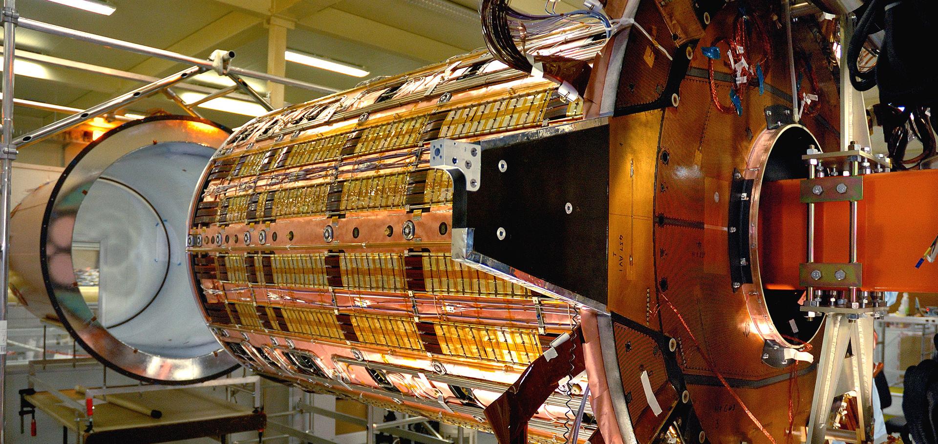
A glimpse into the future of particle therapy
https://physicsworld.com/a/a-glimpse-into-the-future-of-particle-therapy/