
Understanding the challenges of delivering radiotherapy in low- and middle-income countries in Africa
Journal of Cancer Policy Elsevier 35 (2023) 100372
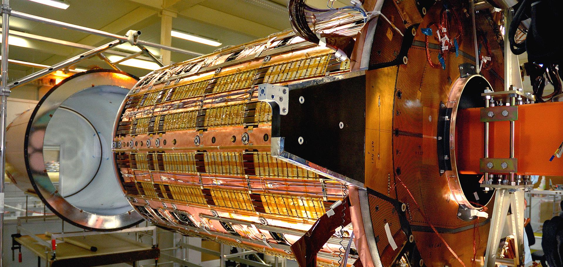
Beam optics study for a potential VHEE beam delivery system
Journal of Physics: Conference Series IOP Publishing 2420:1 (2023)
Abstract:
VHEE (Very High Energy Electron) therapy can be superior to conventional radiotherapy for the treatment of deep seated tumours, whilst not necessarily requiring the space and cost of proton or heavy ion facilities. Developments in high gradient RF technology have allowed electrons to be accelerated to VHEE energies in a compact space, meaning that treatment could be possible with a shorter linac. A crucial component of VHEE treatment is the transfer of the beam from accelerator to patient. This is required to magnify the beam to cover the transverse extent of the tumour, whilst ensuring a uniform beam distribution. Two principle methodologies for the design of a compact transfer line are presented. The first of these is based upon a quadrupole lattice and optical magnification of beam size. A minimisation algorithm is used to enforce certain criteria on the beam distribution at the patient, defining the lattice through an automated routine. Separately, a dual scattering-foil based system is also presented, which uses similar algorithms for the optimisation of the foil geometry in order to achieve the desired beam shape at the patient location.Collaboration: The Force That Makes the Impossible Possible
Advances in Radiation Oncology Elsevier 7:6 (2022) 100966
Clinical use and future requirements of relative biological effectiveness: Survey among all European proton therapy centres
Radiotherapy and Oncology Elsevier 172 (2022) 134-139
Availability of technology for managing cancer patients in the Southeast European (SEE) region
Clinical and Translational Radiation Oncology Elsevier 34 (2022) 57-66