
Radiochromic film dosimetry for VHEE and UHDR: protocol adaptation and verification at the CLEAR facility
Frontiers in Physics Frontiers Media 13 (2025) 1597079
Abstract:
Radiochromic films (RCFs) offer effective two-dimensional dosimetry with a simple, low-cost operating principle, making them suitable for very high-energy electron (VHEE) and ultra-high dose rate (UHDR) applications, where dosimetry standards are lacking. However, achieving high-accuracy measurements with RCFs presents significant challenges, especially in the absence of standardised protocols. To ensure reliable and comparable outcomes, adapted protocols based on a thorough understanding of RCF behaviour are essential. Despite over 6,000 publications addressing RCF protocols, comprehensive guides for high-throughput research machines with small, non-uniform beams are scarce. This paper aims to be a comprehensive guide for non-expert users of RCFs, particularly in VHEE and UHDR research. We identify common errors in RCF preparation, scanning, and processing, proposing strategies to enhance accuracy and efficiency. Using our optimised RCF protocol at the CLEAR facility, we demonstrate a 5% agreement compared to alanine dosimeters irradiated with Gaussian VHEE beams, establishing this protocol as a solid foundation for reliable dosimetry in advanced radiotherapy research.Demonstration and characterisation of a novel dual-scattering system for very high energy electrons
Nuclear Instruments and Methods in Physics Research Section A Accelerators Spectrometers Detectors and Associated Equipment Elsevier (2025) 170943
Abstract:
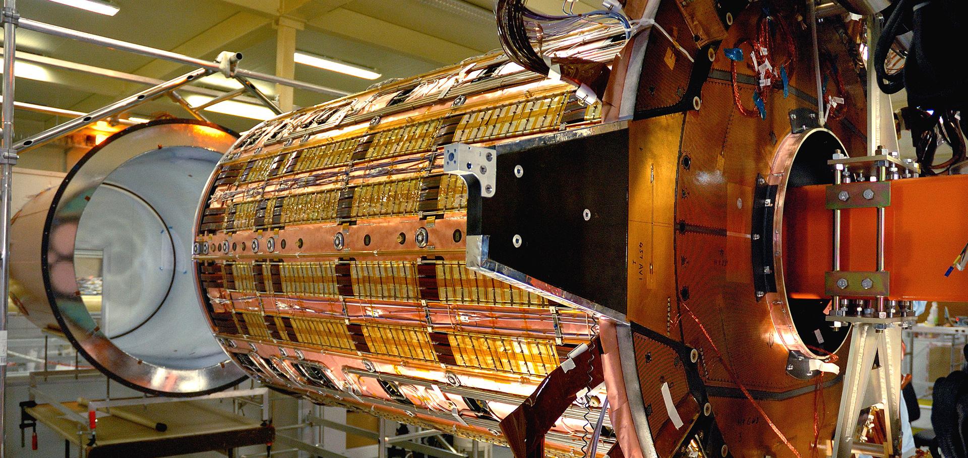
This article describes the design and implementation of the first known prototype of a dual-scattering system for the delivery of a uniform VHEE beam with transverse dimensions suitable for pre-clinical and potential future clinical use. The results presented in this article are the first experimental characterisation of beam flattening with dual-scattering foils in the VHEE regime, confirmed with both profile measurements with a YAG screen and radiochromic EBT3 films. Polylactic acid and aluminium dual-scattering systems were designed using TOPAS Monte-Carlo simulations and multi-objective minimisation methods. Studies to test the success of the scattering systems in providing beam magnification and uniformity were carried out at the 200 MeV CERN Linear Electron Accelerator for Research (CLEAR) facility. A generalised super-Gaussian function was used to model the final beam, and comparisons were made with the simulations used for the design. Transverse profiles with uniform components were measured with each of the scattering systems and quantified with super-Gaussian fitting. The uniformity of the in-air profiles suggested that the superficial dose contributions from X-rays were low. This study demonstrated that magnified VHEE beam profiles with uniform components could be generated and measured at CLEAR. The results from this study were used as a basis for the design of future experiments. Similar systems and design methods could be employed by future clinical VHEE facilities to provide conformal treatment.Modification of the microstructure of the CERN- CLEAR-VHEE beam at the picosecond scale modifies ZFE morphogenesis but has no impact on hydrogen peroxide production
Radiotherapy and Oncology Elsevier (2025) 110942
Multidisciplinary Collaboration and Novel Technological Advances in Hadron Therapy
Technology in Cancer Research & Treatment SAGE Publications 24 (2025) 15330338241311859
Abstract:
The battle against cancer remains a top priority for society, with an urgent need to develop therapies capable of targeting challenging tumours while preserving patient's quality of life. Hadron Therapy (HT), which employs accelerated beams of protons, carbon ions, and other charged particles, represents a significant frontier in cancer treatment. This modality offers superior precision and efficacy compared to conventional methods, delivering therapeutic the dose directly to tumours while sparing healthy tissue. Even though 350,000 patients have already been treated worldwide with protons and 50,000 with carbon ions, HT is still a relatively young field and more research as well as novel, cost-effective and compact accelerator technologies are needed to make this treatment more readily available globally. Interestingly the very first patient was irradiated with protons in September 1954, the same month and year CERN was founded. Both of these endeavours are embedded in cutting edge technologies and multidisciplinary collaboration. HT is finally gaining ground and, even after 70 years, the particle therapy field continues innovating and improving for the benefits of patients globally. Developing technologies that are both affordable and easy to use is key and would allow access to more patients. Advances in accelerator-driven Boron Neutron Capture Therapy (BNCT), image-guided hadron beams delivery, clinical trials and immunotherapy, together with the recent interest and advances in FLASH therapy, which is currently an experimental treatment modality that involves ultrahigh-dose rate delivery, are just a few examples of innovation that may eventually help to provide access to a larger number of patients.Global Collaborations: The European Council for Nuclear Research (CERN) Perspective
Chapter in Global Medical Physics A Guide for International Collaboration, (2025) 171-183