Home
Department Of Physics text logo
  • Research
    • Our research
    • Our research groups
    • Our research in action
    • Research funding support
    • Summer internships for undergraduates
  • Study
    • Undergraduates
    • Postgraduates
  • Engage
    • For alumni
    • For business
    • For schools
    • For the public
Menu
CMP
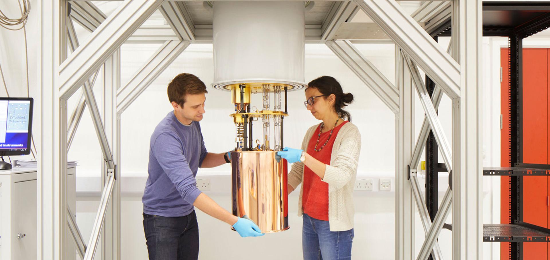
Credit: Jack Hobhouse

Peter Proks

Postdoctoral Research Assistant

Sub department

  • Condensed Matter Physics
peter.proks@physics.ox.ac.uk
Telephone: 72426
Clarendon Laboratory, room 071.4 & 071.7
  • About
  • Publications

Inhibition of the ATP-sensitive potassium channel from mouse pancreatic beta-cells by surfactants.

British journal of pharmacology 124:3 (1998) 529-539

Authors:

PA Smith, P Proks

Abstract:

1. We have used patch-clamp methods to study the effects of the detergents, Cremophor, Tween 80 and Triton X100 on the K(ATP) channel in the pancreatic beta-cell from mouse. 2. All three detergents blocked K(ATP) channel activity with the following order of potency: Tween 80 (Ki< approximately 83 nM)>Triton X100 (Ki=350 nM)>Cremophor. In all cases the block was poorly reversible. 3. Single-channel studies suggested that at low doses, the detergents act as slow blockers of the K(ATP) channel. 4. Unlike the block produced by tolbutamide, that produced by detergent was not affected by intracellular Mg2+-nucleotide, diazoxide or trypsin treatment, nor did it involve an acceleration of rundown or increase in ATP sensitivity of the chanel. 5. The detergents could block the pore-forming subunit, Kir6.2deltaC26, which can be expressed independently of SUR1 (the regulatory subunit of the K(ATP) channel). These data suggest that the detergents act on Kir6.2 and not SUR1. 6. The detergents had no effect on another member of the inward rectifier family: Kir1.1a (ROMK1). 7. Voltage-dependent K-currents in the beta-cell were reversibly blocked by the detergents with a far lower potency than that found for the K(ATP) channel. 8. Like other insulin secretagogues that act by blocking the K(ATP) channel, Cremophor elevated intracellular Ca2+ in single beta-cells to levels that would be expected to elicit insulin secretion. 9. Given the role of the K(ATP) channel in many physiological processes, we conclude that plasma borne detergent may have pharmacological actions mediated through blockage of the K(ATP) channel.
More details from the publisher
More details
More details

Molecular determinants of KATP channel inhibition by ATP.

The EMBO journal 17:12 (1998) 3290-3296

Authors:

SJ Tucker, FM Gribble, P Proks, S Trapp, TJ Ryder, T Haug, F Reimann, FM Ashcroft

Abstract:

ATP-sensitive K+ (KATP) channels are both inhibited and activated by intracellular nucleotides, such as ATP and ADP. The inhibitory effects of nucleotides are mediated via the pore-forming subunit, Kir6.2, whereas the potentiatory effects are conferred by the sulfonylurea receptor subunit, SUR. The stimulatory action of Mg-nucleotides complicates analysis of nucleotide inhibition of Kir6. 2/SUR1 channels. We therefore used a truncated isoform of Kir6.2, that expresses ATP-sensitive channels in the absence of SUR1, to explore the mechanism of nucleotide inhibition. We found that Kir6.2 is highly selective for ATP, and that both the adenine moiety and the beta-phosphate contribute to specificity. We also identified several mutations that significantly reduce ATP inhibition. These are located in two distinct regions of Kir6.2: the N-terminus preceding, and the C-terminus immediately following, the transmembrane domains. Some mutations in the C-terminus also markedly increased the channel open probability, which may account for the decrease in apparent ATP sensitivity. Other mutations did not affect the single-channel kinetics, and may reduce ATP inhibition by interfering with ATP binding and/or the link between ATP binding and pore closure. Our results also implicate the proximal C-terminus in KATP channel gating.
More details from the publisher
More details

Electrophysiological and metabolic characterization of single beta-cells and islets from diabetic GK rats.

Diabetes 47:1 (1998) 73-81

Authors:

SJ Hughes, M Faehling, CW Thorneley, P Proks, FM Ashcroft, PA Smith

Abstract:

We have used the whole-cell recording technique to determine whether ATP-sensitive potassium (K[ATP]) currents, voltage-dependent Ca2+ currents, and exocytosis are different in single beta-cells from pancreatic islets of Goto-Kakizaki (GK) rats, a novel model of NIDDM, and normal rats. In addition, we have also measured the insulin secretory responses, ATP content, and the rate of glucose metabolism in intact islets. Although the glucose sensitivity of the K(ATP) current was similar between GK rats and controls, in the absence of glucose, K(ATP) current density was larger in GK rats, which resulted in a more hyperpolarized membrane potential. Whole-cell Ca2+ currents were similar. By monitoring the cell capacitance with a fixed intracellular solution, no difference was detected in the exocytotic responses of beta-cells from normal and GK rats. In islets from GK rats, the rates of glucose utilization ([3H]H2O production from 5-[3H]glucose) and oxidation ([14C]CO2 production from U-[14C]glucose) were not significantly different from controls. Insulin secretion, however, was impaired (by 50%), and this was paralleled by a smaller increase in ATP content in response to stimulation by 10 mmol/l glucose in islets from GK rats when compared with controls. Under conditions in which K(ATP) channels were held open and the effects of glucose were independent of membrane potential, insulin release was still significantly lower in GK rat islets than in controls. These findings suggest that the impaired insulin secretion in islets from GK rats does not simply result from a failure to close K(ATP) channels, nor does it result from an impairment in calcium secretion coupling.
More details from the publisher
More details
More details

Phentolamine block of KATP channels is mediated by Kir6.2.

Proceedings of the National Academy of Sciences of the United States of America 94:21 (1997) 11716-11720

Authors:

P Proks, FM Ashcroft

Abstract:

The ATP-sensitive K+-channel (KATP channel) plays a key role in insulin secretion from pancreatic beta cells. It is closed both by glucose metabolism and the sulfonylurea drugs that are used in the treatment of noninsulin-dependent diabetes mellitus, thereby initiating a membrane depolarization that activates voltage-dependent Ca2+ entry and insulin release. The beta cell KATP channel is a complex of two proteins: Kir6.2 and SUR1. The former is an ATP-sensitive K+-selective pore, whereas SUR1 is a channel regulator that endows Kir6.2 with sensitivity to sulfonylureas. A number of drugs containing an imidazoline moiety, such as phentolamine, also act as potent stimulators of insulin secretion, but their mechanism of action is unknown. We have used a truncated form of Kir6.2, which expresses independently of SUR1, to show that phentolamine does not inhibit KATP channels by interacting with SUR1. Instead, our results argue that phentolamine may interact directly with Kir6.2 to produce a voltage-independent reduction in channel activity. The single-channel conductance is unaffected. Although the ATP molecule also contains an imidazoline group, the site at which phentolamine blocks is not identical to the ATP-inhibitory site, because phentolamine block of an ATP-insensitive mutant (K185Q) is normal. KATP channels also are found in the heart where they are involved in the response to cardiac ischemia: they also are blocked by phentolamine. Our results suggest that this may be because Kir6.2, which is expressed in the heart, forms the pore of the cardiac KATP channel.
More details from the publisher
More details
More details

Rapid ATP-dependent priming of secretory granules precedes Ca(2+)-induced exocytosis in mouse pancreatic B-cells.

The Journal of physiology 503 ( Pt 2) (1997) 399-412

Authors:

L Eliasson, E Renström, WG Ding, P Proks, P Rorsman

Abstract:

1. The glucose and ATP dependence of exocytosis were investigated in single mouse pancreatic B-cells by monitoring changes in cell capacitance evoked by voltage-clamp depolarizations, infusion of high [Ca2+]i buffers or photorelease of caged Ca2+ or ATP. 2. In intact B-cells, using the perforated patch whole-cell technique, glucose (5 mM) increased exocytotic responses evoked by membrane depolarization 5-fold over that observed in the absence of the sugar. Increasing the glucose concentration to 20 mM produced a further doubling of exocytosis. The stimulatory action of glucose was attributable to glucose metabolism and abolished by mannoheptulose, an inhibitor of glucose phosphorylation. 3. Exocytosis triggered by infusion of high [Ca2+]i and ATP was reduced by 80% when ATP was replaced by its non-hydrolysable analogue adenosine 5'-[beta, gamma-methylene]triphosphate (AMP-PCP) in standard whole-cell experiments. Exocytosis elicited by GTP gamma S was similarly affected by replacement of ATP with the stable analogue. 4. Photoreleasing ATP in the presence of 170 nM [Ca2+]i, following the complete wash-out of endogenous ATP produced a prompt (latency, < 400 ms) and biphasic stimulation of exocytosis. 5. Elevation of [Ca2+]i to exocytotic levels by photorelease from Ca(2+)-nitrophenyl EGTA preloaded into the cell evoked a biphasic stimulation in the presence of Mg-ATP. The response consisted of an initial rapid (completed in < 200 ms) phase followed by a slower (lasting > or = 10 s) sustained component. Replacement of ATP with AMP-PCP abolished the late component but did not affect the initial phase. The latency between elevation of [Ca2+]i and exocytosis was determined as < 45 ms. Inclusion of N-ethylmaleimide (NEM; 0.5 mM for 3 min) in the intracellular solution exerted effects similar to those obtained by substituting AMP-PCP for ATP. 6. We conclude that the B-cell contains a small pool (40 granules) of primed granules which are immediately available for release and which are capable of undergoing exocytosis in an ATP-independent fashion. We propose that this pool of granules is preferentially released during first phase glucose-stimulated insulin secretion. The short latency between the application of ATP and the onset of exocytosis finally suggests that priming takes place with sufficient speed to participate in the rapid adjustment of the secretory capacity of the B-cell.
More details from the publisher
More details
More details

Pagination

  • First page First
  • Previous page Prev
  • …
  • Page 17
  • Page 18
  • Page 19
  • Page 20
  • Current page 21
  • Page 22
  • Page 23
  • Page 24
  • Page 25
  • Next page Next
  • Last page Last

Footer Menu

  • Contact us
  • Giving to the Dept of Physics
  • Work with us
  • Media

User account menu

  • Log in

Follow us

FIND US

Clarendon Laboratory,

Parks Road,

Oxford,

OX1 3PU

CONTACT US

Tel: +44(0)1865272200

University of Oxfrod logo Department Of Physics text logo
IOP Juno Champion logo Athena Swan Silver Award logo

© University of Oxford - Department of Physics

Cookies | Privacy policy | Accessibility statement

Built by: Versantus

  • Home
  • Research
  • Study
  • Engage
  • Our people
  • News & Comment
  • Events
  • Our facilities & services
  • About us
  • Current students
  • Staff intranet