Home
Department Of Physics text logo
  • Research
    • Our research
    • Our research groups
    • Our research in action
    • Research funding support
    • Summer internships for undergraduates
  • Study
    • Undergraduates
    • Postgraduates
  • Engage
    • For alumni
    • For business
    • For schools
    • For the public
Menu
CMP
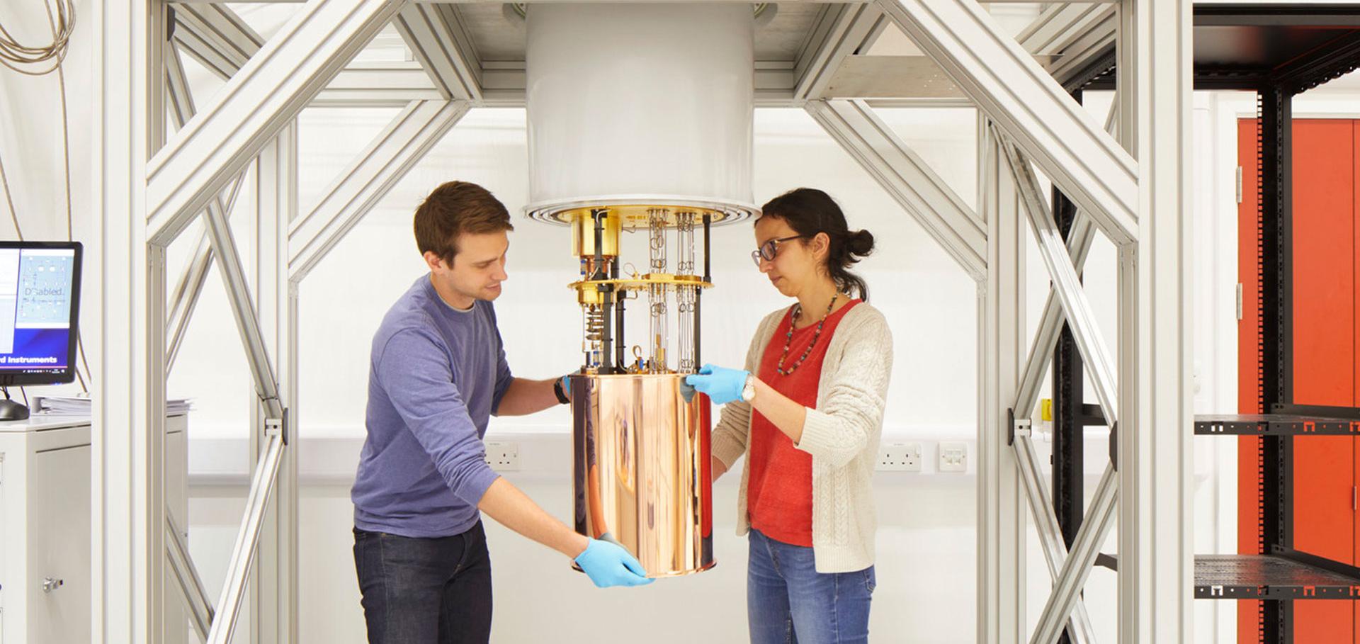
Credit: Jack Hobhouse

Professor Achillefs Kapanidis

Professor of Biological Physics

Research theme

  • Biological physics

Sub department

  • Condensed Matter Physics

Research groups

  • Gene machines
Achillefs.Kapanidis@physics.ox.ac.uk
Telephone: 01865 (2)72226
Biochemistry Building
groups.physics.ox.ac.uk/genemachines/group
  • About
  • Publications

Internalization and observation of fluorescent biomolecules in living microorganisms via electroporation.

Journal of visualized experiments : JoVE (2015)

Authors:

Louise Aigrain, Marko Sustarsic, Robert Crawford, Anne Plochowietz, Achillefs N Kapanidis

Abstract:

The ability to study biomolecules in vivo is crucial for understanding their function in a biological context. One powerful approach involves fusing molecules of interest to fluorescent proteins such as GFP to study their expression, localization and function. However, GFP and its derivatives are significantly larger and less photostable than organic fluorophores generally used for in vitro experiments, and this can limit the scope of investigation. We recently introduced a straightforward, versatile and high-throughput method based on electroporation, allowing the internalization of biomolecules labeled with organic fluorophores into living microorganisms. Here we describe how to use electroporation to internalize labeled DNA fragments or proteins into Escherichia coli and Saccharomyces cerevisiæ, how to quantify the number of internalized molecules using fluorescence microscopy, and how to quantify the viability of electroporated cells. Data can be acquired at the single-cell or single-molecule level using fluorescence or FRET. The possibility of internalizing non-labeled molecules that trigger a physiological observable response in vivo is also presented. Finally, strategies of optimization of the protocol for specific biological systems are discussed.
More details from the publisher
More details
More details

Real-time transcription initiation by E. coli RNA polymerase in vitro and in vivo

Biophysical Journal Cell Press 108:2, Supplement 1 (2015) 115A

Authors:

Anne Plochowietz, Diego Duchi Llumigusin, Pawel Zawadzki, Afaf H El-Sagheer, Thomas Brown, Achillefs N Kapanidis
More details from the publisher
Details from ORA
More details

Single-Molecule FRET for Dynamic Structural Biology: DNA Polymerase i Structure and Mechanism with Angstrom Precision

Biophysical Journal Elsevier 108:2 (2015) 14a

Authors:

Marko Sustarsic, Timothy Craggs, Johannes Hohlbein, Andrew Cuthbert, Nicholas Taylor, Hendrik Kaju, Majid Mosayebi, Jonathan Doye, Achillefs N Kapanidis
More details from the publisher

Studies of the FtsK DNA Translocase using Two-Color Tethered Fluorophore Motion

Biophysical Journal Elsevier 108:2 (2015) 67a

Authors:

Peter FJ May, Pawel Zawadzki, Lidia K Arciszewska, David Sherratt, Achillefs N Kapanidis
More details from the publisher

In vivo single-molecule imaging of bacterial DNA replication, transcription, and repair.

FEBS letters 588:19 (2014) 3585-3594

Authors:

Mathew Stracy, Stephan Uphoff, Federico Garza de Leon, Achillefs N Kapanidis

Abstract:

In vivo single-molecule experiments offer new perspectives on the behaviour of DNA binding proteins, from the molecular level to the length scale of whole bacterial cells. With technological advances in instrumentation and data analysis, fluorescence microscopy can detect single molecules in live cells, opening the doors to directly follow individual proteins binding to DNA in real time. In this review, we describe key technical considerations for implementing in vivo single-molecule fluorescence microscopy. We discuss how single-molecule tracking and quantitative super-resolution microscopy can be adapted to extract DNA binding kinetics, spatial distributions, and copy numbers of proteins, as well as stoichiometries of protein complexes. We highlight experiments which have exploited these techniques to answer important questions in the field of bacterial gene regulation and transcription, as well as chromosome replication, organisation and repair. Together, these studies demonstrate how single-molecule imaging is transforming our understanding of DNA-binding proteins in cells.
More details from the publisher
More details
More details

Pagination

  • First page First
  • Previous page Prev
  • …
  • Page 18
  • Page 19
  • Page 20
  • Page 21
  • Current page 22
  • Page 23
  • Page 24
  • Page 25
  • Page 26
  • …
  • Next page Next
  • Last page Last

Footer Menu

  • Contact us
  • Giving to the Dept of Physics
  • Work with us
  • Media

User account menu

  • Log in

Follow us

FIND US

Clarendon Laboratory,

Parks Road,

Oxford,

OX1 3PU

CONTACT US

Tel: +44(0)1865272200

University of Oxfrod logo Department Of Physics text logo
IOP Juno Champion logo Athena Swan Silver Award logo

© University of Oxford - Department of Physics

Cookies | Privacy policy | Accessibility statement

Built by: Versantus

  • Home
  • Research
  • Study
  • Engage
  • Our people
  • News & Comment
  • Events
  • Our facilities & services
  • About us
  • Current students
  • Staff intranet