Home
Department Of Physics text logo
  • Research
    • Our research
    • Our research groups
    • Our research in action
    • Research funding support
    • Summer internships for undergraduates
  • Study
    • Undergraduates
    • Postgraduates
  • Engage
    • For alumni
    • For business
    • For schools
    • For the public
Menu
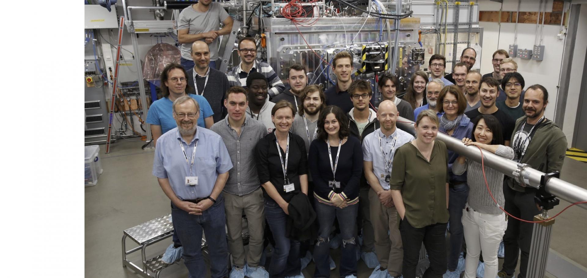
First HED experiment at XFEL

Professor Justin Wark

Professor of Physics

Sub department

  • Atomic and Laser Physics

Research groups

  • Oxford Centre for High Energy Density Science (OxCHEDS)
Justin.Wark@physics.ox.ac.uk
Telephone: 01865 (2)72251
Clarendon Laboratory, room 029.9
  • About
  • Publications

Simulations of in situ x-ray diffraction from uniaxially compressed highly textured polycrystalline targets

Journal of Applied Physics AIP Publishing 118:6 (2015) 065902

Authors:

David McGonegle, Despina Milathianaki, Bruce A Remington, Justin S Wark, Andrew Higginbotham
More details from the publisher
More details

Direct Observation of Melting in Shock-Compressed Bismuth With Femtosecond X-ray Diffraction.

Physical review letters 115:9 (2015) 095701

Authors:

MG Gorman, R Briggs, EE McBride, A Higginbotham, B Arnold, JH Eggert, DE Fratanduono, E Galtier, AE Lazicki, HJ Lee, HP Liermann, B Nagler, A Rothkirch, RF Smith, DC Swift, GW Collins, JS Wark, MI McMahon

Abstract:

The melting of bismuth in response to shock compression has been studied using in situ femtosecond x-ray diffraction at an x-ray free electron laser. Both solid-solid and solid-liquid phase transitions are documented using changes in discrete diffraction peaks and the emergence of broad, liquid scattering upon release from shock pressures up to 14 GPa. The transformation from the solid state to the liquid is found to occur in less than 3 ns, very much faster than previously believed. These results are the first quantitative measurements of a liquid material obtained on shock release using x-ray diffraction, and provide an upper limit for the time scale of melting of bismuth under shock loading.
More details from the publisher
More details
More details

Imaging Shock Waves in Diamond with Both High Temporal and Spatial Resolution at an XFEL.

Scientific reports 5 (2015) 11089

Authors:

Andreas Schropp, Robert Hoppe, Vivienne Meier, Jens Patommel, Frank Seiboth, Yuan Ping, Damien G Hicks, Martha A Beckwith, Gilbert W Collins, Andrew Higginbotham, Justin S Wark, Hae Ja Lee, Bob Nagler, Eric C Galtier, Brice Arnold, Ulf Zastrau, Jerome B Hastings, Christian G Schroer

Abstract:

The advent of hard x-ray free-electron lasers (XFELs) has opened up a variety of scientific opportunities in areas as diverse as atomic physics, plasma physics, nonlinear optics in the x-ray range, and protein crystallography. In this article, we access a new field of science by measuring quantitatively the local bulk properties and dynamics of matter under extreme conditions, in this case by using the short XFEL pulse to image an elastic compression wave in diamond. The elastic wave was initiated by an intense optical laser pulse and was imaged at different delay times after the optical pump pulse using magnified x-ray phase-contrast imaging. The temporal evolution of the shock wave can be monitored, yielding detailed information on shock dynamics, such as the shock velocity, the shock front width, and the local compression of the material. The method provides a quantitative perspective on the state of matter in extreme conditions.
More details from the publisher
More details
More details

Imaging transient melting of a nanocrystal using an X-ray laser.

Proceedings of the National Academy of Sciences of the United States of America 112:24 (2015) 7444-7448

Authors:

Jesse N Clark, Loren Beitra, Gang Xiong, David M Fritz, Henrik T Lemke, Diling Zhu, Matthieu Chollet, Garth J Williams, Marc M Messerschmidt, Brian Abbey, Ross J Harder, Alexander M Korsunsky, Justin S Wark, David A Reis, Ian K Robinson

Abstract:

There is a fundamental interest in studying photoinduced dynamics in nanoparticles and nanostructures as it provides insight into their mechanical and thermal properties out of equilibrium and during phase transitions. Nanoparticles can display significantly different properties from the bulk, which is due to the interplay between their size, morphology, crystallinity, defect concentration, and surface properties. Particularly interesting scenarios arise when nanoparticles undergo phase transitions, such as melting induced by an optical laser. Current theoretical evidence suggests that nanoparticles can undergo reversible nonhomogenous melting with the formation of a core-shell structure consisting of a liquid outer layer. To date, studies from ensembles of nanoparticles have tentatively suggested that such mechanisms are present. Here we demonstrate imaging transient melting and softening of the acoustic phonon modes of an individual gold nanocrystal, using an X-ray free electron laser. The results demonstrate that the transient melting is reversible and nonhomogenous, consistent with a core-shell model of melting. The results have implications for understanding transient processes in nanoparticles and determining their elastic properties as they undergo phase transitions.
More details from the publisher
More details
More details

Single Hit Energy-resolved Laue Diffraction.

The Review of scientific instruments 86:5 (2015) 053908

Authors:

Shamim Patel, Matthew J Suggit, Paul G Stubley, James A Hawreliak, Orlando Ciricosta, Andrew J Comley, Gilbert W Collins, Jon H Eggert, John M Foster, Justin S Wark, Andrew Higginbotham

Abstract:

In situ white light Laue diffraction has been successfully used to interrogate the structure of single crystal materials undergoing rapid (nanosecond) dynamic compression up to megabar pressures. However, information on strain state accessible via this technique is limited, reducing its applicability for a range of applications. We present an extension to the existing Laue diffraction platform in which we record the photon energy of a subset of diffraction peaks. This allows for a measurement of the longitudinal and transverse strains in situ during compression. Consequently, we demonstrate measurement of volumetric compression of the unit cell, in addition to the limited aspect ratio information accessible in conventional white light Laue. We present preliminary results for silicon, where only an elastic strain is observed. VISAR measurements show the presence of a two wave structure and measurements show that material downstream of the second wave does not contribute to the observed diffraction peaks, supporting the idea that this material may be highly disordered, or has undergone large scale rotation.
More details from the publisher
More details
More details
Details from ArXiV

Pagination

  • First page First
  • Previous page Prev
  • …
  • Page 17
  • Page 18
  • Page 19
  • Page 20
  • Current page 21
  • Page 22
  • Page 23
  • Page 24
  • Page 25
  • …
  • Next page Next
  • Last page Last

Footer Menu

  • Contact us
  • Giving to the Dept of Physics
  • Work with us
  • Media

User account menu

  • Log in

Follow us

FIND US

Clarendon Laboratory,

Parks Road,

Oxford,

OX1 3PU

CONTACT US

Tel: +44(0)1865272200

University of Oxfrod logo Department Of Physics text logo
IOP Juno Champion logo Athena Swan Silver Award logo

© University of Oxford - Department of Physics

Cookies | Privacy policy | Accessibility statement

Built by: Versantus

  • Home
  • Research
  • Study
  • Engage
  • Our people
  • News & Comment
  • Events
  • Our facilities & services
  • About us
  • Current students
  • Staff intranet