Home
Department Of Physics text logo
  • Research
    • Our research
    • Our research groups
    • Our research in action
    • Research funding support
    • Summer internships for undergraduates
  • Study
    • Undergraduates
    • Postgraduates
  • Engage
    • For alumni
    • For business
    • For schools
    • For the public
Menu
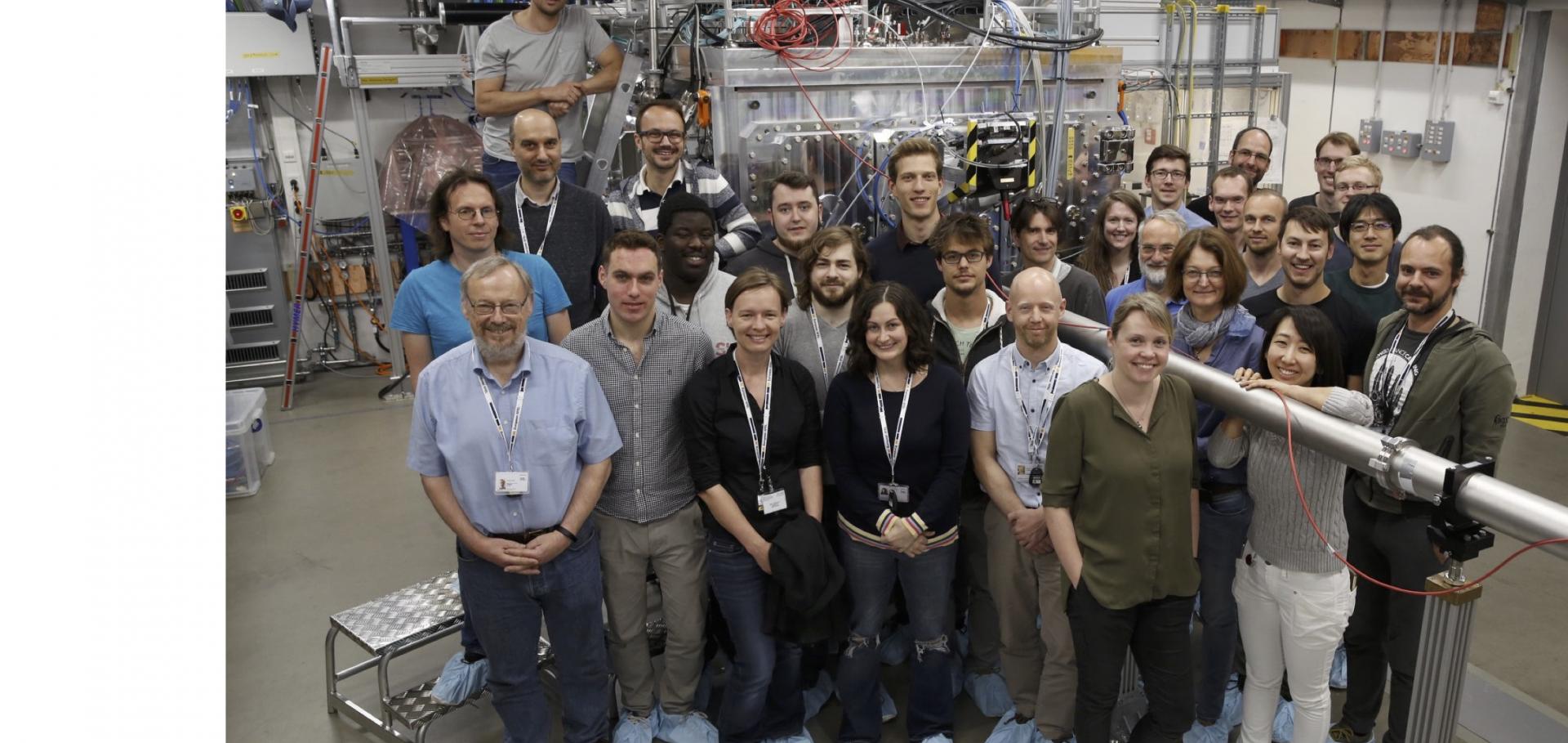
First HED experiment at XFEL

Professor Justin Wark

Professor of Physics

Sub department

  • Atomic and Laser Physics

Research groups

  • Oxford Centre for High Energy Density Science (OxCHEDS)
Justin.Wark@physics.ox.ac.uk
Telephone: 01865 (2)72251
Clarendon Laboratory, room 029.9
  • About
  • Publications

High-quality ultra-fast total scattering and pair distribution function data using an X-ray free-electron laser.

IUCrJ International Union of Crystallography (IUCr) 12:5 (2025)

Authors:

Adam F Sapnik, Philip A Chater, Dean S Keeble, John SO Evans, Federica Bertolotti, Antonietta Guagliardi, Lise J Støckler, Elodie A Harbourne, Anders B Borup, Rebecca S Silberg, Adrien Descamps, Clemens Prescher, Benjamin D Klee, Axel Phelipeau, Imran Ullah, Kárel G Medina, Tobias A Bird, Viktoria Kaznelson, William Lynn, Andrew L Goodwin, Bo B Iversen, Celine Crepisson, Emil S Bozin, Kirsten MØ Jensen, Emma E McBride, Reinhard B Neder, Ian Robinson, Justin S Wark, Michał Andrzejewski, Ulrike Boesenberg, Erik Brambrink, Carolina Camarda, Valerio Cerantola, Sebastian Goede, Hauke Höppner, Oliver S Humphries, Zuzana Konopkova, Naresh Kujala, Thomas Michelat, Motoaki Nakatsutsumi, Alexander Pelka, Thomas R Preston, Lisa Randolph, Michael Roeper, Andreas Schmidt, Cornelius Strohm, Minxue Tang, Peter Talkovski, Ulf Zastrau, Karen Appel, David A Keen

Abstract:

High-quality total scattering data, a key tool for understanding atomic-scale structure in disordered materials, require stable instrumentation and access to high momentum transfers. This is now routine at dedicated synchrotron instrumentation using high-energy X-ray beams, but it is very challenging to measure a total scattering dataset in less than a few microseconds. This limits their effectiveness for capturing structural changes that occur at the much faster timescales of atomic motion. Current X-ray free-electron lasers (XFELs) provide femtosecond-pulsed X-ray beams with maximum energies of ∼24 keV, giving the potential to measure total scattering and the attendant pair distribution functions (PDFs) on femtosecond timescales. We demonstrate that this potential has been realized using the HED scientific instrument at the European XFEL and present normalized total scattering data for 0.35 Å-1 < Q < 16.6 Å-1 and their PDFs from a broad spectrum of materials, including crystalline, nanocrystalline and amorphous solids, liquids and clusters in solution. We analyzed the data using a variety of methods, including Rietveld refinement, small-box PDF refinement, joint reciprocal-real-space refinement, cluster refinement and Debye scattering analysis. The resolution function of the setup is also characterized. We conclusively show that high-quality data can be obtained from a single ∼30 fs XFEL pulse for multiple different sample types. Our efforts not only significantly increase the existing maximum reported Q range for an S(Q) measured at an XFEL but also mean that XFELs are now a viable X-ray source for the broad community of people using reciprocal-space total scattering and PDF methods in their research.
More details from the publisher
More details

High-quality ultra-fast total scattering and pair distribution function data using an X-ray free electron laser

IUCrJ International Union of Crystallography 12:5 (2025) 12

Authors:

Adam F Sapnik, Philip A Chater, Dean S Keeble, Elodie Harbourne, Andrew Goodwin, Celine Crepisson, Justin Wark

Abstract:

High-quality total scattering data, a key tool for understanding atomic-scale structure in disordered materials, require stable instrumentation and access to high momentum transfers. This is now routine at dedicated synchrotron instrumentation using high-energy X-ray beams, but it is very challenging to measure a total scattering dataset in less than a few microseconds. This limits their effectiveness for capturing structural changes that occur at the much faster timescales of atomic motion. Current X-ray free-electron lasers (XFELs) provide femtosecond-pulsed X-ray beams with maximum energies of ~24 keV, giving the potential to measure total scattering and the attendant pair distribution functions (PDFs) on femtosecond timescales. Here, we show that this potential has been realised using the HED scientific instrument at the European XFEL and present normalised total scattering data for 0.35Å−1 < Q < 16.6Å−1 and their PDFs from a broad spectrum of materials, including crystalline, nanocrystalline and amorphous solids, liquids, and clusters in solution. We analyse the data using a variety of methods, including Rietveld refinement, small-box PDF refinement, joint reciprocal–real space refinement, cluster refinement, and Debye scattering analysis. The resolution function of the setup is also characterised. We conclusively show that high-quality data can be obtained from a single ~30 fs XFEL pulse for multiple different sample types. Our efforts not only significantly increase the existing maximum reported Q-range for an S(Q) measured at an XFEL but also mean that XFELs are now a viable X-ray source for the broad community of people using reciprocal space total scattering and PDF methods in their research.
More details from the publisher
Details from ORA

Calibration and characterization of the line-VISAR diagnostic at the HED-HIBEF instrument at the European XFEL

Review of Scientific Instruments AIP Publishing 96:7 (2025) 075206

Authors:

A Descamps, TM Hutchinson, R Briggs, EE McBride, M Millot, T Michelat, JH Eggert, B Albertazzi, L Antonelli, MR Armstrong, C Baehtz, OB Ball, S Banerjee, AB Belonoshko, A Benuzzi-Mounaix, CA Bolme, V Bouffetier, K Buakor, T Butcher, V Cerantola, J Chantel, AL Coleman, J Collier, G Collins, AJ Comley, F Coppari, TE Cowan, C Crépisson, G Cristoforetti, H Cynn, S Di Dio Cafiso, F Dorchies, MJ Duff, A Dwivedi, D Errandonea, E Galtier, H Ginestet, L Gizzi, A Gleason, S Goede, JM Gonzalez, MG Gorman, M Harmand, NJ Hartley, PG Heighway, C Hernandez-Gomez, A Higginbotham, H Höppner, RJ Husband, H Hwang, J Kim, P Koester, Z Konopkova, D Kraus, A Krygier, L Labate, A Laso Garcia, AE Lazicki, Y Lee, P Mason, M Masruri, B Massani, D McGonegle, C McGuire, JD McHardy, RS McWilliams, S Merkel, G Morard, B Nagler, M Nakatsutsumi, K Nguyen-Cong, A-M Norton, II Oleynik, C Otzen, N Ozaki, S Pandolfi, DJ Peake, A Pelka, KA Pereira, JP Phillips, C Prescher, TR Preston, L Randolph, D Ranjan, A Ravasio, R Redmer, J Rips, D Santamaria-Perez, DJ Savage, M Schoelmerich, J-P Schwinkendorf, S Singh, J Smith, RF Smith, A Sollier, J Spear, C Spindloe, M Stevenson, C Strohm, T-A Suer, M Tang, T Tschentscher, M Toncian, T Toncian, SJ Tracy, M Tyldesley, CE Vennari, T Vinci, TJ Volz, J Vorberger, JPS Walsh, JS Wark, JT Willman, L Wollenweber, U Zastrau, E Brambrink, K Appel, MI McMahon

Abstract:

In dynamic-compression experiments, the line-imaging Velocity Interferometer System for Any Reflector (VISAR) is a well-established diagnostic used to probe the velocity history, including wave profiles derived from dynamically compressed interfaces and wavefronts, depending on material optical properties. Knowledge of the velocity history allows for the determination of the pressure achieved during compression. Such a VISAR analysis is often based on Fourier transform techniques and assumes that the recorded interferograms are free from image distortions. In this paper, we describe the VISAR diagnostic installed at the HED-HIBEF instrument located at the European XFEL along with its calibration and characterization. It comprises a two-color (532, 1064 nm), three-arm (with three velocity sensitivities) line imaging system. We provide a procedure to correct VISAR images for geometric distortions and evaluate the performance of the system using Fourier analysis. We finally discuss the spatial and temporal calibrations of the diagnostic. As an example, we compare the pressure extracted from the VISAR analysis of shock-compressed polyimide and silicon.
More details from the publisher
More details

The structure of liquid carbon elucidated by in situ X-ray diffraction

Nature Nature Research 642:8067 (2025) 351-355

Authors:

D Kraus, J Rips, M Schörner, MG Stevenson, J Vorberger, D Ranjan, J Lütgert, B Heuser, JH Eggert, H-P Liermann, II Oleynik, S Pandolfi, R Redmer, A Sollier, C Strohm, TJ Volz, B Albertazzi, SJ Ali, L Antonelli, C Bähtz, OB Ball, S Banerjee, AB Belonoshko, CA Bolme

Abstract:

Carbon has a central role in biology and organic chemistry, and its solid allotropes provide the basis of much of our modern technology1. However, the liquid form of carbon remains nearly uncharted2, and the structure of liquid carbon and most of its physical properties are essentially unknown3. But liquid carbon is relevant for modelling planetary interiors4, 5 and the atmospheres of white dwarfs6, as an intermediate state for the synthesis of advanced carbon materials7, 8, inertial confinement fusion implosions9, hypervelocity impact events on carbon materials10 and our general understanding of structured fluids at extreme conditions11. Here we present a precise structure measurement of liquid carbon at pressures of around 1 million atmospheres obtained by in situ X-ray diffraction at an X-ray free-electron laser. Our results show a complex fluid with transient bonding and approximately four nearest neighbours on average, in agreement with quantum molecular dynamics simulations. The obtained data substantiate the understanding of the liquid state of one of the most abundant elements in the universe and can test models of the melting line. The demonstrated experimental abilities open the path to performing similar studies of the structure of liquids composed of light elements at extreme conditions.
More details from the publisher
Details from ORA
More details
More details

Isostructural phase transition of Fe2O3 under laser shock compression

Physical Review Letters American Physical Society 134:17 (2025) 176102

Authors:

Alexis Amouretti, Celine Crepisson, Sam Azadi, Francois Brisset, Delphine Cabaret, Thomas Campbell, David Chin, Gilbert Rip Collins, Linda Hansen, Guillaume Fiquet, Alessandro Forte, Thomas Gawne, Francois Guyot, Patrick Heighway, Eva Heripre, Eric Cunningham, Hae Ja Lee, David McGonegle, Bob Nagler, Juan Pintor, Danae Polsin, Gaelle Rousse, Yuanfeng Shi, Ethan Smith, Justin Wark, Sam Vinko, Marion Harmand

Abstract:

We present in situ x-ray diffraction and velocity measurements of Fe2⁢O3 under laser shock compression at pressures between 38–122 GPa. None of the high-pressure phases reported by static compression studies were observed. Instead, we observed an isostructural phase transition from 𝛼−Fe2⁢O3 to a new 𝛼′−Fe2⁢O3 phase at a pressure of 50–62 GPa. The 𝛼′−Fe2⁢O3 phase differs from 𝛼−Fe2⁢O3 by an 11% volume drop and a different unit cell compressibility. We further observed a two-wave structure in the velocity profile, which can be related to an intermediate regime where both 𝛼 and 𝛼′ phases coexist. Density functional theory calculations with a Hubbard parameter indicate that the observed unit cell volume drop can be associated with a spin transition following a magnetic collapse.
More details from the publisher
Details from ORA
More details
More details

Pagination

  • Current page 1
  • Page 2
  • Page 3
  • Page 4
  • Page 5
  • Page 6
  • Page 7
  • Page 8
  • Page 9
  • …
  • Next page Next
  • Last page Last

Footer Menu

  • Contact us
  • Giving to the Dept of Physics
  • Work with us
  • Media

User account menu

  • Log in

Follow us

FIND US

Clarendon Laboratory,

Parks Road,

Oxford,

OX1 3PU

CONTACT US

Tel: +44(0)1865272200

University of Oxfrod logo Department Of Physics text logo
IOP Juno Champion logo Athena Swan Silver Award logo

© University of Oxford - Department of Physics

Cookies | Privacy policy | Accessibility statement

Built by: Versantus

  • Home
  • Research
  • Study
  • Engage
  • Our people
  • News & Comment
  • Events
  • Our facilities & services
  • About us
  • Current students
  • Staff intranet