Preparing high purity initial states for nuclear magnetic resonance quantum computing
Physical Review Letters 93:4 (2004) 1-40501
Abstract:
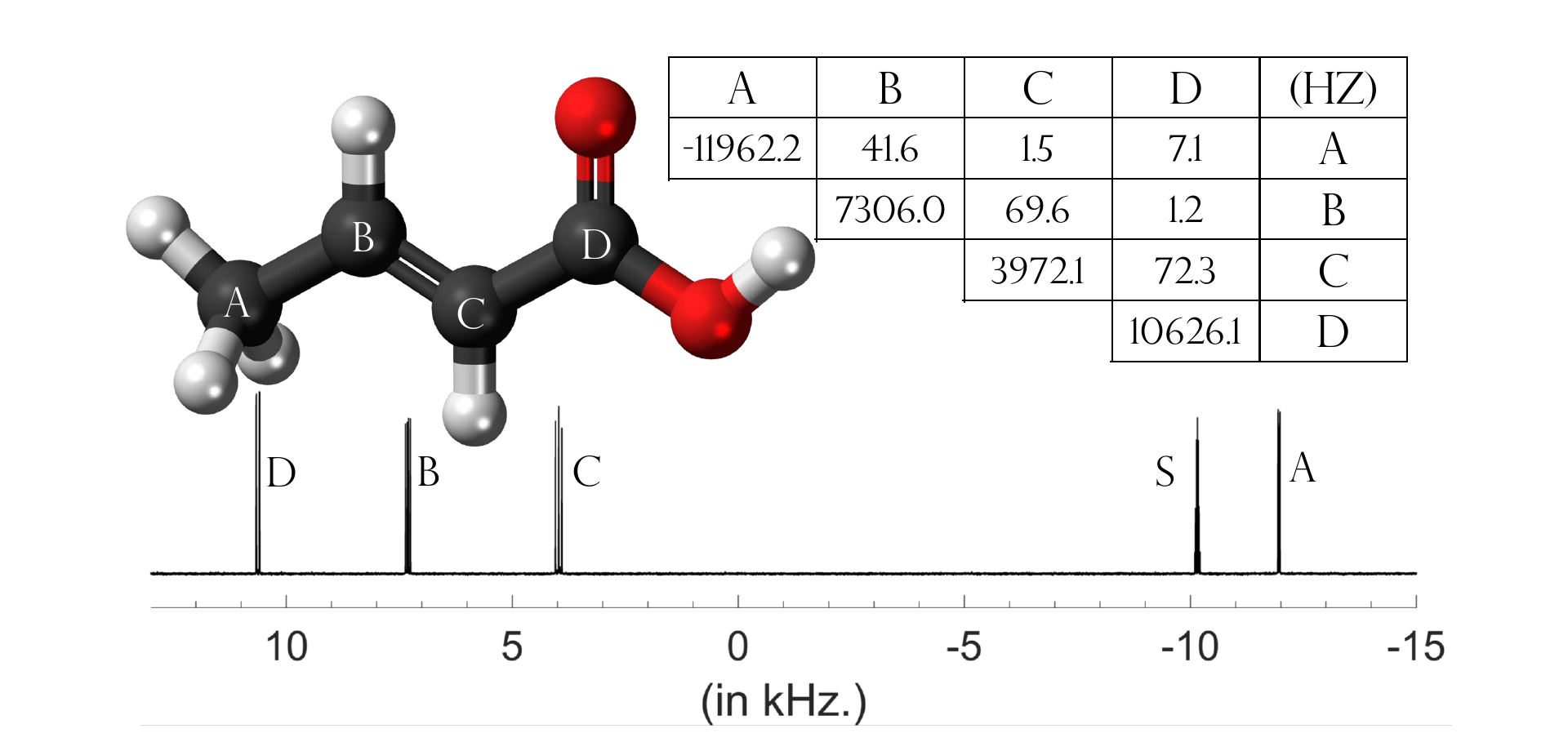
High purity initial states were prepared for nuclear magnetic resonance quantum computing. A chemical reaction involving pure parahydrogen was initiated using a 12 ns laser pulse. The product containes a hydrogen-derived two-spin system with an effective spin-state purity of 0.916. It is observed that the resulting spin state has an entanglement of formation of 0.822 and cannot be described by local hidden variable models.Implementing Grover's Quantum Search on a Para-Hydrogen based Pure State NMR Quantum Computer
(2004)
Implementation of NMR quantum computation with para-hydrogen derived high purity quantum states
(2004)
A combined parahydrogen and theoretical study of H2 activation by 16-electron d8 ruthenium(0) complexes and their subsequent catalytic behaviour
DALTON TRANSACTIONS (2004) 3616-3628
Course 10 Nuclear magnetic resonance quantum computation
Les Houches - Ecole d'Ete de Physique Theorique Elsevier 79 (2004) 357-400