Home
Department Of Physics text logo
  • Research
    • Our research
    • Our research groups
    • Our research in action
    • Research funding support
    • Summer internships for undergraduates
  • Study
    • Undergraduates
    • Postgraduates
  • Engage
    • For alumni
    • For business
    • For schools
    • For the public
Menu
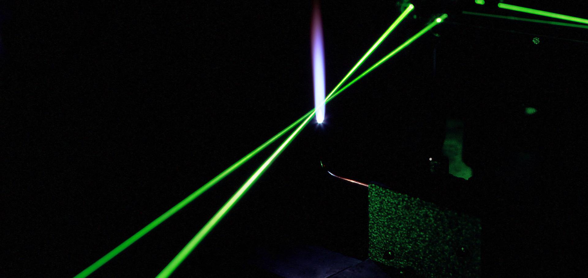
Lasers in flame

Probing temperature and radical species in a flame using 4-wave mixing spectroscopy.

Professor Paul Ewart

Emeritus Professor

Sub department

  • Atomic and Laser Physics
paul.ewart@physics.ox.ac.uk
  • About
  • Publications

Multi-species detection using multi-mode absorption spectroscopy (MUMAS)

Applied Physics B: Lasers and Optics 111:4 (2013) 627-635

Authors:

JH Northern, AWJ Thompson, ML Hamilton, P Ewart

Abstract:

The detection of multiple species using a single laser and single detector employing multi-mode absorption spectroscopy (MUMAS) is reported. An in-house constructed, diode-pumped, Er:Yb:glass micro-laser operating at 1,565 nm with 10 modes separated by 18 GHz was used to record MUMAS signals in a gas mixture containing C2H2, N2O and CO. The components of the mixture were detected simultaneously by identifying multiple transitions in each of the species. By using temperature- and pressure-dependent modelled spectral fits to the data, partial pressures of each species in the mixture were determined with an uncertainty of ±2 %. © 2013 Springer-Verlag Berlin Heidelberg.
More details from the publisher

Multi-species detection using multi-mode absorption spectroscopy (MUMAS)

Applied Physics B: Lasers and Optics (2013) 1-9

Authors:

JH Northern, AWJ Thompson, ML Hamilton, P Ewart

Abstract:

The detection of multiple species using a single laser and single detector employing multi-mode absorption spectroscopy (MUMAS) is reported. An in-house constructed, diode-pumped, Er:Yb:glass micro-laser operating at 1,565 nm with 10 modes separated by 18 GHz was used to record MUMAS signals in a gas mixture containing CH, NO and CO. The components of the mixture were detected simultaneously by identifying multiple transitions in each of the species. By using temperature- and pressure-dependent modelled spectral fits to the data, partial pressures of each species in the mixture were determined with an uncertainty of ±2 %. © 2013 Springer-Verlag Berlin Heidelberg.
More details from the publisher
More details

X

Callaloo Johns Hopkins University Press 36:4 (2013) 850-850

Authors:

Édouard Glissant, Mary Ann Caws
More details from the publisher

Cardiac electrophysiological imaging systems scalable for high-throughput drug testing

Pflugers Archiv European Journal of Physiology 464:6 (2012) 645-656

Authors:

P Lee, K Wang, CE Woods, P Yan, P Kohl, P Ewart, LM Loew, DA Terrar, C Bollensdorff

Abstract:

Multi-parametric electrophysiological measurements using optical methods have become a highly valued standard in cardiac research. Most published optical mapping systems are expensive and complex. Although some applications demand high-cost components and complex designs, many can be tackled with simpler solutions. Here, we describe (1) a camera-based voltage and calcium imaging system using a single 'economy' electron-multiplying charge-coupled device camera and demonstrate the possibility of using a consumer camera for imaging calcium transients of the heart, and (2) a photodiode-based voltage and calcium high temporal resolution measurement system using single-element photodiodes and an optical fibre. High-throughput drug testing represents an application where system scalability is particularly attractive. Therefore, we tested our systems on tissue exposed to a well-characterized and clinically relevant calcium channel blocker, nifedipine, which has been used to treat angina and hypertension. As experimental models, we used the Langendorff-perfused whole-heart and thin ventricular tissue slices, a preparation gaining renewed interest by the cardiac research community. Using our simplified systems, we were able to monitor simultaneously the marked changes in the voltage and calcium transients that are responsible for the negative inotropic effect of the compound.
More details from the publisher

Cardiac electrophysiological imaging systems scalable for high-throughput drug testing.

Pflugers Arch 464:6 (2012) 645-656

Authors:

Peter Lee, Ken Wang, Christopher E Woods, Ping Yan, Peter Kohl, Paul Ewart, Leslie M Loew, Derek A Terrar, Christian Bollensdorff

Abstract:

Multi-parametric electrophysiological measurements using optical methods have become a highly valued standard in cardiac research. Most published optical mapping systems are expensive and complex. Although some applications demand high-cost components and complex designs, many can be tackled with simpler solutions. Here, we describe (1) a camera-based voltage and calcium imaging system using a single 'economy' electron-multiplying charge-coupled device camera and demonstrate the possibility of using a consumer camera for imaging calcium transients of the heart, and (2) a photodiode-based voltage and calcium high temporal resolution measurement system using single-element photodiodes and an optical fibre. High-throughput drug testing represents an application where system scalability is particularly attractive. Therefore, we tested our systems on tissue exposed to a well-characterized and clinically relevant calcium channel blocker, nifedipine, which has been used to treat angina and hypertension. As experimental models, we used the Langendorff-perfused whole-heart and thin ventricular tissue slices, a preparation gaining renewed interest by the cardiac research community. Using our simplified systems, we were able to monitor simultaneously the marked changes in the voltage and calcium transients that are responsible for the negative inotropic effect of the compound.
More details from the publisher
More details

Pagination

  • First page First
  • Previous page Prev
  • …
  • Page 4
  • Page 5
  • Page 6
  • Page 7
  • Current page 8
  • Page 9
  • Page 10
  • Page 11
  • Page 12
  • …
  • Next page Next
  • Last page Last

Footer Menu

  • Contact us
  • Giving to the Dept of Physics
  • Work with us
  • Media

User account menu

  • Log in

Follow us

FIND US

Clarendon Laboratory,

Parks Road,

Oxford,

OX1 3PU

CONTACT US

Tel: +44(0)1865272200

University of Oxfrod logo Department Of Physics text logo
IOP Juno Champion logo Athena Swan Silver Award logo

© University of Oxford - Department of Physics

Cookies | Privacy policy | Accessibility statement

Built by: Versantus

  • Home
  • Research
  • Study
  • Engage
  • Our people
  • News & Comment
  • Events
  • Our facilities & services
  • About us
  • Current students
  • Staff intranet