
Cardiac electrophysiological imaging systems scalable for high-throughput drug testing.
Pflugers Arch 464:6 (2012) 645-656
Abstract:
Multi-parametric electrophysiological measurements using optical methods have become a highly valued standard in cardiac research. Most published optical mapping systems are expensive and complex. Although some applications demand high-cost components and complex designs, many can be tackled with simpler solutions. Here, we describe (1) a camera-based voltage and calcium imaging system using a single 'economy' electron-multiplying charge-coupled device camera and demonstrate the possibility of using a consumer camera for imaging calcium transients of the heart, and (2) a photodiode-based voltage and calcium high temporal resolution measurement system using single-element photodiodes and an optical fibre. High-throughput drug testing represents an application where system scalability is particularly attractive. Therefore, we tested our systems on tissue exposed to a well-characterized and clinically relevant calcium channel blocker, nifedipine, which has been used to treat angina and hypertension. As experimental models, we used the Langendorff-perfused whole-heart and thin ventricular tissue slices, a preparation gaining renewed interest by the cardiac research community. Using our simplified systems, we were able to monitor simultaneously the marked changes in the voltage and calcium transients that are responsible for the negative inotropic effect of the compound.Simultaneous measurement and modulation of multiple physiological parameters in the isolated heart using optical techniques
Pflugers Archiv European Journal of Physiology 464:4 (2012) 403-414
Abstract:
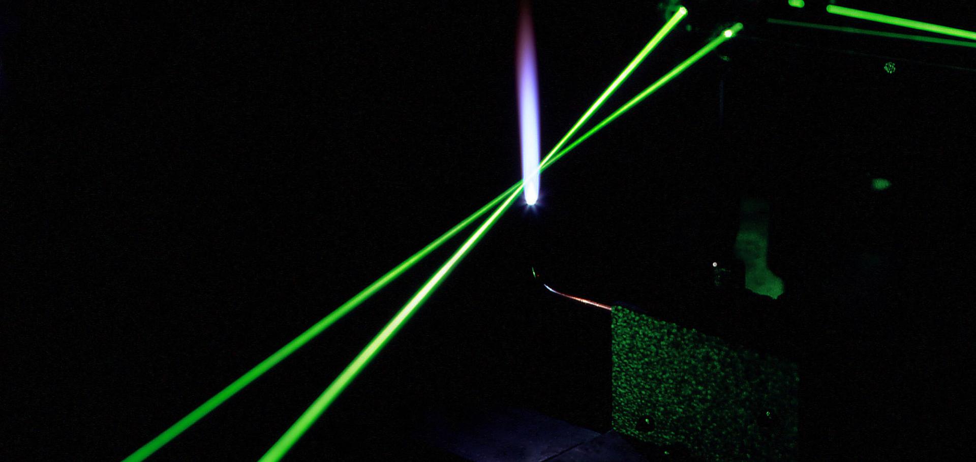
Whole-heart multi-parametric optical mapping has provided valuable insight into the interplay of electrophysiological parameters, and this technology will continue to thrive as dyes are improved and technical solutions for imaging become simpler and cheaper. Here, we show the advantage of using improved 2nd-generation voltage dyes, provide a simple solution to panoramic multi-parametric mapping, and illustrate the application of flash photolysis of caged compounds for studies in the whole heart. For proof of principle, we used the isolated rat whole-heart model. After characterising the blue and green isosbestic points of di-4-ANBDQBS and di-4-ANBDQPQ, respectively, two voltage and calcium mapping systems are described. With two newly custom-made multi-band optical filters, (1) di-4-ANBDQBS and fluo-4 and (2) di-4-ANBDQPQ and rhod-2 mapping are demonstrated. Furthermore, we demonstrate three-parameter mapping using di-4-ANBDQPQ, rhod-2 and NADH. Using off-the-shelf optics and the di-4- ANBDQPQ and rhod-2 combination, we demonstrate panoramic multi-parametric mapping, affording a 360° spatiotemporal record of activity. Finally, local optical perturbation of calcium dynamics in the whole heart is demonstrated using the caged compound, o-nitrophenyl ethylene glycol tetraacetic acid (NP-EGTA), with an ultraviolet light-emitting diode (LED). Calcium maps (heart loaded with di-4-ANBDQPQ and rhod-2) demonstrate successful NP-EGTA loading and local flash photolysis. All imaging systems were built using only a single camera. In conclusion, using novel 2nd-generation voltage dyes, we developed scalable techniques for multi-parametric optical mapping of the whole heart from one point of view and panoramically. In addition to these parameter imaging approaches, we show that it is possible to use caged compounds and ultraviolet LEDs to locally perturb electrophysiological parameters in the whole heart. © The Author(s) 2012.Simultaneous measurement and modulation of multiple physiological parameters in the isolated heart using optical techniques.
Pflugers Arch 464:4 (2012) 403-414
Abstract:
Whole-heart multi-parametric optical mapping has provided valuable insight into the interplay of electrophysiological parameters, and this technology will continue to thrive as dyes are improved and technical solutions for imaging become simpler and cheaper. Here, we show the advantage of using improved 2nd-generation voltage dyes, provide a simple solution to panoramic multi-parametric mapping, and illustrate the application of flash photolysis of caged compounds for studies in the whole heart. For proof of principle, we used the isolated rat whole-heart model. After characterising the blue and green isosbestic points of di-4-ANBDQBS and di-4-ANBDQPQ, respectively, two voltage and calcium mapping systems are described. With two newly custom-made multi-band optical filters, (1) di-4-ANBDQBS and fluo-4 and (2) di-4-ANBDQPQ and rhod-2 mapping are demonstrated. Furthermore, we demonstrate three-parameter mapping using di-4-ANBDQPQ, rhod-2 and NADH. Using off-the-shelf optics and the di-4-ANBDQPQ and rhod-2 combination, we demonstrate panoramic multi-parametric mapping, affording a 360° spatiotemporal record of activity. Finally, local optical perturbation of calcium dynamics in the whole heart is demonstrated using the caged compound, o-nitrophenyl ethylene glycol tetraacetic acid (NP-EGTA), with an ultraviolet light-emitting diode (LED). Calcium maps (heart loaded with di-4-ANBDQPQ and rhod-2) demonstrate successful NP-EGTA loading and local flash photolysis. All imaging systems were built using only a single camera. In conclusion, using novel 2nd-generation voltage dyes, we developed scalable techniques for multi-parametric optical mapping of the whole heart from one point of view and panoramically. In addition to these parameter imaging approaches, we show that it is possible to use caged compounds and ultraviolet LEDs to locally perturb electrophysiological parameters in the whole heart.Simultaneous voltage and calcium mapping of genetically purified human induced pluripotent stem cell-derived cardiac myocyte monolayers.
Circ Res 110:12 (2012) 1556-1563
Abstract:
RATIONALE: Human induced pluripotent stem cell-derived cardiomyocytes (iPSC-CMs) offer a powerful in vitro tool to investigate disease mechanisms and to perform patient-specific drug screening. To date, electrophysiological analysis of iPSC-CMs has been limited to single-cell recordings or low-resolution microelectrode array mapping of small cardiomyocyte aggregates. New methods of generating and optically mapping impulse propagation of large human iPSC-CM cardiac monolayers are needed. OBJECTIVE: Our first aim was to develop an imaging platform with versatility for multiparameter electrophysiological mapping of cardiac preparations, including human iPSC-CM monolayers. Our second aim was to create large electrically coupled human iPSC-CM monolayers for simultaneous action potential and calcium wave propagation measurements. METHODS AND RESULTS: A fluorescence imaging platform based on electronically controlled light-emitting diode illumination, a multiband emission filter, and single camera sensor was developed and utilized to monitor simultaneously action potential and intracellular calcium wave propagation in cardiac preparations. Multiple, large-diameter (≥1 cm), electrically coupled human cardiac monolayers were then generated that propagated action potentials and calcium waves at velocities similar to those commonly observed in rodent cardiac monolayers. CONCLUSIONS: The multiparametric imaging system presented here offers a scalable enabling technology to measure simultaneously action potential and intracellular calcium wave amplitude and dynamics of cardiac monolayers. The advent of large-scale production of human iPSC-CMs makes it possible to now generate sufficient numbers of uniform cardiac monolayers that can be utilized for the study of arrhythmia mechanisms and offers advantages over commonly used rodent models.Flash Photolysis of Caged Compounds during Simultaneous Imaging of Calcium and Voltage in the Whole Heart using Light-Emitting-Diodes
BIOPHYSICAL JOURNAL 102:3 (2012) 671A-671A