Home
Department Of Physics text logo
  • Research
    • Our research
    • Our research groups
    • Our research in action
    • Research funding support
    • Summer internships for undergraduates
  • Study
    • Undergraduates
    • Postgraduates
  • Engage
    • For young people
    • For teachers
    • For the public
    • For alumni
    • For business
  • Support
Menu
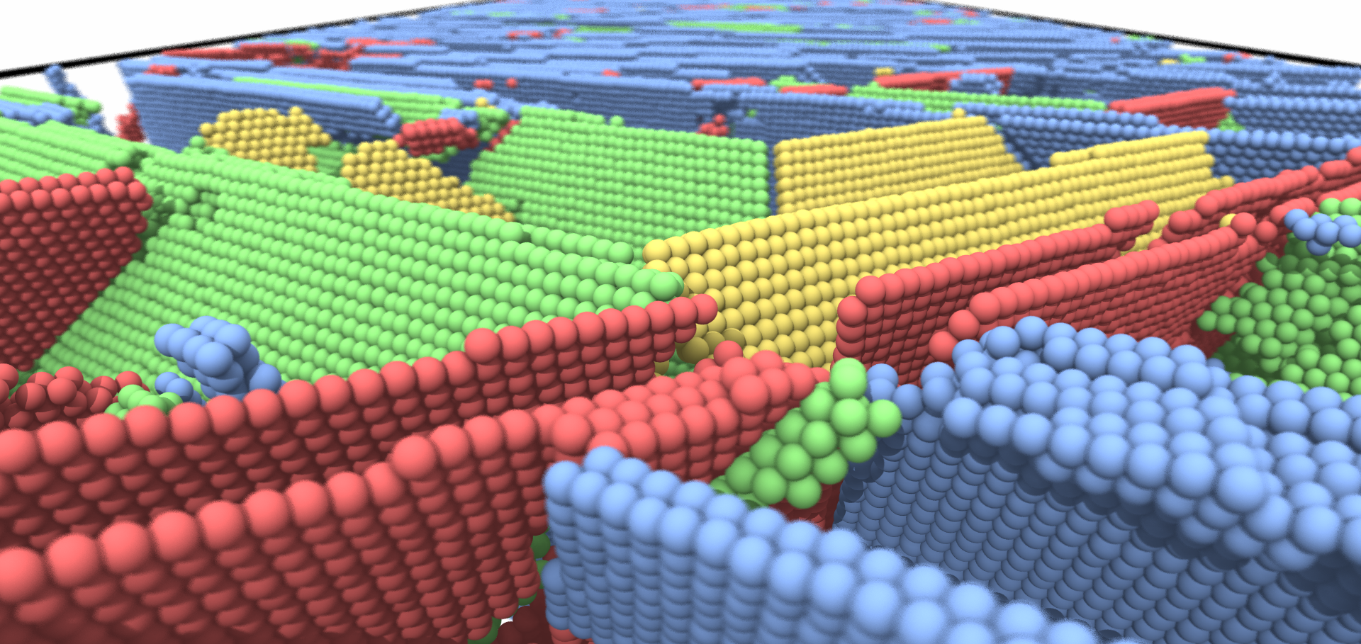
Stacking faults in shock-compressed copper

Shock-induced stacking faults in dynamically compressed copper modelled using large-scale molecular dynamics simulations

Dr Paddy Heighway

Early Career OxCHEDS Research Fellow

Research theme

  • Lasers and high energy density science

Sub department

  • Atomic and Laser Physics

Research groups

  • Oxford Centre for High Energy Density Science (OxCHEDS)
patrick.heighway@physics.ox.ac.uk
Clarendon Laboratory, room Simon
  • About
  • Publications

Featured Work

Molecular dynamics simulation of double-slip in a bcc crystal
Giving the slip to a metal deformation mystery

A kinematic model uses X-ray diffraction patterns to identify active slip systems during the dynamic compression of metals

Link to Scilight

Ultrafast temperature diagnosis of dynamically compressed matter using millielectronvolt inelastic x-ray scattering beyond the first Brillouin zone

Journal of Applied Physics AIP Publishing 140:3 (2026) 35903

Authors:

Pg Heighway, Js Wark

Abstract:

<jats:p>We present calculations of the millielectronvolt-scale x-ray scattering spectra of multilayered dynamic-compression targets comprising an unstructured ablator layer and a crystalline, textured sample layer. Our model builds on the classic formulation of x-ray thermal diffuse scattering by Warren [Acta Crystallogr. 6, 803 (1953)] and includes both elastic and first-order (single-phonon) inelastic scattering contributions to the dynamic structure factor S(q, ω). We focus on the umklapp scattering regime (i.e., at momentum transfers outside the first Brillouin zone) where the ablator scattering that threatens to overwhelm the inelastic scattering from the crystalline layer of interest is suppressed. We show that, despite the considerably more complex structure of the inelastic scattering spectra in this intermediate-q regime, it is still possible to reliably deduce the temperature of the crystal using Dornheim’s Laplace-transform–based formalism [Dornheim et al., Phys. Plasmas 30, 042707 (2023)], regardless of the details of the sample’s texture.</jats:p>
More details from the publisher
More details

Structural evolution of iron oxides melts at Earth’s outer-core pressures

Nature Communications Springer Nature (2026)

Authors:

Céline Crépisson, Mila Fitzgerald, Domenic Peake, Patrick G Heighway, Thomas Stevens, Adrien Descamps, David McGonegle, Alexis Amouretti, Karim K Alaa El-Din, Michal Andrzejewski, Sam Azadi, Erik Brambrink, Carolina Camarda, David A Chin, Samuele Di Dio Cafiso, Ana Coutinho Dutra, Hauke Höppner, Kohdai Yamamoto, Phani S Karamched, Zuzana Konôpková, Motoaki Nakatsutsumi, Norimasa Ozaki, Danae N Polsin, Jan-Patrick Schwinkendorf, Georgiy Shoulga, Cornelius Strohm, Minxue Tang, Harry Taylor, Monika Toncian, Yizhen Wang, Jin Yao, Gianluca Gregori, Justin S Wark, Karen Appel, Marion Harmand, Sam M Vinko

Abstract:

Oxygen and other light elements comprise up to 5 wt% of the Earth’s outer-core, and may significantly influence its physical properties and the operation of the geodynamo. Here we report in situ X-ray diffraction measurements of Fe, Fe + 4.5 FeO (atomic proportion), and Fe2O3 melts at 177-440 GPa, achieved using laser-driven shock compression at an x-ray free-electron laser. The melts exhibit Fe-O coordination numbers between 4.0(0.4) and 4.5(0.4), indicating predominantly four-fold coordination environments. These coordination states are significantly smaller than those of Fe-bearing lower-mantle phases such as bridgmanite and ferropericlase. Shorter Fe-Fe interatomic distances in compressed iron oxide melts drive the denser packing relative to ambient melts, while the structural differences between Fe + 4.5 FeO and Fe2O3 melts under shock indicate that the oxidation state modulates oxygen solubility in liquid Fe. At 177 GPa ( ~ 380 km below the core-mantle boundary) and 3800 K, Fe2O3 melts exhibit higher Fe-O coordination, suggesting that local variations in oxygen content could contribute to the stratification in the uppermost outer-core inferred from seismological and geomagnetic observations.
More details from the publisher
More details
More details

X-ray thermal diffuse scattering as a texture-robust temperature diagnostic for dynamically compressed solids

Journal of Applied Physics AIP Publishing 138:15 (2025) 155903

Authors:

Pg Heighway, Dj Peake, T Stevens, Js Wark, B Albertazzi, Sj Ali, L Antonelli, Mr Armstrong, C Baehtz, Ob Ball, S Banerjee, Ab Belonoshko, Ca Bolme, V Bouffetier, R Briggs, K Buakor, T Butcher, S Di Dio Cafiso, V Cerantola, J Chantel, A Di Cicco, Al Coleman, J Collier, G Collins, Aj Comley, F Coppari, Te Cowan, G Cristoforetti, H Cynn, A Descamps, F Dorchies, Mj Duff, A Dwivedi, C Edwards, Jh Eggert, D Errandonea, G Fiquet, E Galtier, A Laso Garcia, H Ginestet, L Gizzi, A Gleason, S Goede, Jm Gonzalez, Mg Gorman, M Harmand, Nj Hartley, C Hernandez-Gomez, A Higginbotham, H Höppner

Abstract:

We present a model of x-ray thermal diffuse scattering (TDS) from a cubic polycrystal with an arbitrary crystallographic texture, based on the classic approach of Warren [B. E. Warren, Acta Crystallogr. 6, 803 (1953)]. We compare the predictions of our model with femtosecond x-ray diffraction patterns gathered from ambient and dynamically compressed rolled copper foils obtained at the High Energy Density instrument of the European X-Ray Free-Electron Laser facility and find that the texture-aware TDS model yields more accurate results than does the conventional powder model owed to Warren. Nevertheless, we further show: with sufficient angular detector coverage, the TDS signal is largely unchanged by sample orientation and in all cases strongly resembles the signal from a perfectly random powder; shot-to-shot fluctuations in the TDS signal resulting from grain-sampling statistics are at the percent level, in stark contrast to the fluctuations in the Bragg-peak intensities (which are over an order of magnitude greater); and TDS is largely unchanged even following texture evolution caused by compression-induced plastic deformation. We conclude that TDS is robust against texture variation, making it a flexible temperature diagnostic applicable just as well to off-the-shelf commercial foils as to ideal powders.
More details from the publisher
More details

X-ray thermal diffuse scattering as a texture-robust temperature diagnostic for dynamically compressed solids

(2025)

Authors:

PG Heighway, DJ Peake, T Stevens, JS Wark, B Albertazzi, SJ Ali, L Antonelli, MR Armstrong, C Baehtz, OB Ball, S Banerjee, AB Belonoshko, CA Bolme, V Bouffetier, R Briggs, K Buakor, T Butcher, S Di Dio Cafiso, V Cerantola, J Chantel, A Di Cicco, AL Coleman, J Collier, G Collins, AJ Comley, F Coppari, TE Cowan, G Cristoforetti, H Cynn, A Descamps, F Dorchies, MJ Duff, A Dwivedi, C Edwards, JH Eggert, D Errandonea, G Fiquet, E Galtier, A Laso Garcia, H Ginestet, L Gizzi, A Gleason, S Goede, JM Gonzalez, MG Gorman, M Harmand, N Hartley, C Hernandez-Gomez, A Higginbotham, H Höppner, OS Humphries, RJ Husband, TM Hutchinson, H Hwang, DA Keen, J Kim, P Koester, Z Konopkova, D Kraus, A Krygier, L Labate, AE Lazicki, Y Lee, H-P Liermann, P Mason, M Masruri, B Massani, EE McBride, C McGuire, JD McHardy, D McGonegle, RS McWilliams, S Merkel, G Morard, B Nagler, M Nakatsutsumi, K Nguyen-Cong, A-M Norton, II Oleynik, C Otzen, N Ozaki, S Pandolfi, A Pelka, KA Pereira, JP Phillips, C Prescher, T Preston, L Randolph, D Ranjan, A Ravasio, J Rips, D Santamaria-Perez, DJ Savage, M Schoelmerich, J-P Schwinkendorf, S Singh, J Smith, RF Smith, A Sollier, J Spear, C Spindloe, M Stevenson, C Strohm, T-A Suer, M Tang, M Toncian, T Toncian, SJ Tracy, A Trapananti, T Tschentscher, M Tyldesley, CE Vennari, T Vinci, SC Vogel, TJ Volz, J Vorberger, JT Willman, L Wollenweber, U Zastrau, E Brambrink, K Appel, MI McMahon
Details from ArXiV

Calibration and characterization of the line-VISAR diagnostic at the HED-HIBEF instrument at the European XFEL

Review of Scientific Instruments AIP Publishing 96:7 (2025) 075206

Authors:

A Descamps, TM Hutchinson, R Briggs, EE McBride, M Millot, T Michelat, JH Eggert, B Albertazzi, L Antonelli, MR Armstrong, C Baehtz, OB Ball, S Banerjee, AB Belonoshko, A Benuzzi-Mounaix, CA Bolme, V Bouffetier, K Buakor, T Butcher, V Cerantola, J Chantel, AL Coleman, J Collier, G Collins, AJ Comley, F Coppari, TE Cowan, C Crépisson, G Cristoforetti, H Cynn, S Di Dio Cafiso, F Dorchies, MJ Duff, A Dwivedi, D Errandonea, E Galtier, H Ginestet, L Gizzi, A Gleason, S Goede, JM Gonzalez, MG Gorman, M Harmand, NJ Hartley, PG Heighway, C Hernandez-Gomez, A Higginbotham, H Höppner, RJ Husband, H Hwang, J Kim, P Koester, Z Konopkova, D Kraus, A Krygier, L Labate, A Laso Garcia, AE Lazicki, Y Lee, P Mason, M Masruri, B Massani, D McGonegle, C McGuire, JD McHardy, RS McWilliams, S Merkel, G Morard, B Nagler, M Nakatsutsumi, K Nguyen-Cong, A-M Norton, II Oleynik, C Otzen, N Ozaki, S Pandolfi, DJ Peake, A Pelka, KA Pereira, JP Phillips, C Prescher, TR Preston, L Randolph, D Ranjan, A Ravasio, R Redmer, J Rips, D Santamaria-Perez, DJ Savage, M Schoelmerich, J-P Schwinkendorf, S Singh, J Smith, RF Smith, A Sollier, J Spear, C Spindloe, M Stevenson, C Strohm, T-A Suer, M Tang, T Tschentscher, M Toncian, T Toncian, SJ Tracy, M Tyldesley, CE Vennari, T Vinci, TJ Volz, J Vorberger, JPS Walsh, JS Wark, JT Willman, L Wollenweber, U Zastrau, E Brambrink, K Appel, MI McMahon

Abstract:

In dynamic-compression experiments, the line-imaging Velocity Interferometer System for Any Reflector (VISAR) is a well-established diagnostic used to probe the velocity history, including wave profiles derived from dynamically compressed interfaces and wavefronts, depending on material optical properties. Knowledge of the velocity history allows for the determination of the pressure achieved during compression. Such a VISAR analysis is often based on Fourier transform techniques and assumes that the recorded interferograms are free from image distortions. In this paper, we describe the VISAR diagnostic installed at the HED-HIBEF instrument located at the European XFEL along with its calibration and characterization. It comprises a two-color (532, 1064 nm), three-arm (with three velocity sensitivities) line imaging system. We provide a procedure to correct VISAR images for geometric distortions and evaluate the performance of the system using Fourier analysis. We finally discuss the spatial and temporal calibrations of the diagnostic. As an example, we compare the pressure extracted from the VISAR analysis of shock-compressed polyimide and silicon.
More details from the publisher
More details

Pagination

  • First page First
  • Previous page Prev
  • Page 1
  • Current page 2
  • Page 3
  • Page 4
  • Page 5
  • Page 6
  • Page 7
  • Next page Next
  • Last page Last

Footer Menu

  • Contact us
  • Giving to the Dept of Physics
  • Work with us
  • Media

User account menu

  • Log in

Follow us

FIND US

Clarendon Laboratory,

Parks Road,

Oxford,

OX1 3PU

CONTACT US

Tel: +44(0)1865272200

University of Oxfrod logo Department Of Physics text logo
IOP Juno Champion logo Athena Swan Silver Award logo

© University of Oxford - Department of Physics

Cookies | Privacy policy | Accessibility statement

Built by: Versantus

  • Home
  • Research
  • Study
  • Engage
  • Our people
  • News & Comment
  • Events
  • Our facilities & services
  • About us
  • Giving to Physics
  • Current students
  • Staff intranet