Home
Department Of Physics text logo
  • Research
    • Our research
    • Our research groups
    • Our research in action
    • Research funding support
    • Summer internships for undergraduates
  • Study
    • Undergraduates
    • Postgraduates
  • Engage
    • For alumni
    • For business
    • For schools
    • For the public
Menu
CMP
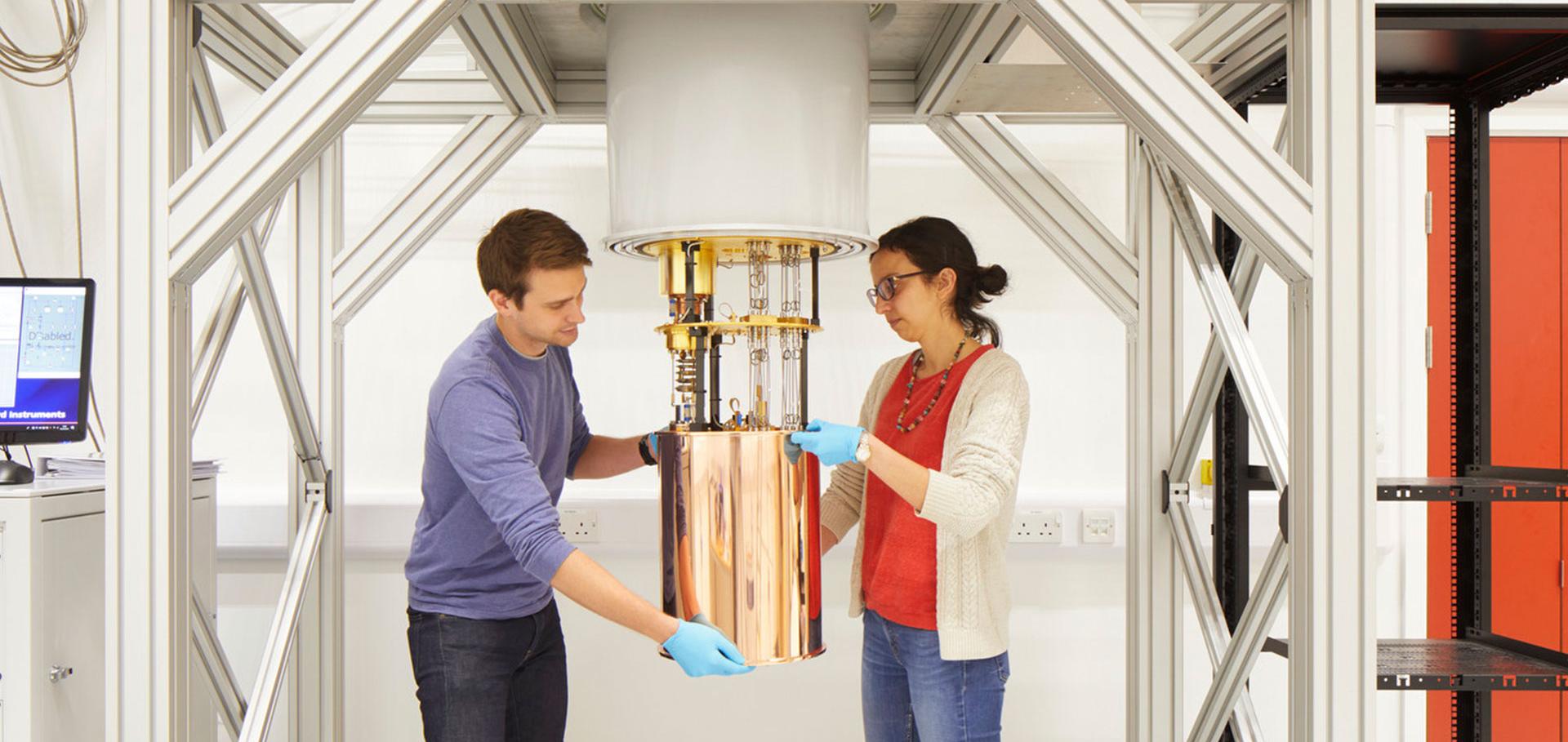
Credit: Jack Hobhouse

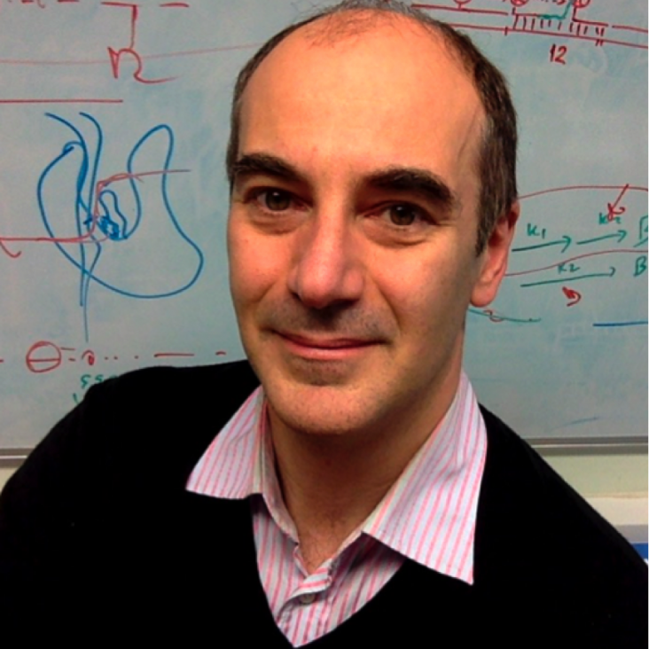
Professor Achillefs Kapanidis

Professor of Biological Physics

Research theme

  • Biological physics

Sub department

  • Condensed Matter Physics

Research groups

  • Gene machines
Achillefs.Kapanidis@physics.ox.ac.uk
Telephone: 01865 (2)72226
Biochemistry Building
groups.physics.ox.ac.uk/genemachines/group
  • About
  • Publications

Capturing reaction paths and intermediates in Cre-loxP recombination using single-molecule fluorescence.

Proc Natl Acad Sci U S A 109:51 (2012) 20871-20876

Authors:

Justin NM Pinkney, Pawel Zawadzki, Jaroslaw Mazuryk, Lidia K Arciszewska, David J Sherratt, Achillefs N Kapanidis

Abstract:

Site-specific recombination plays key roles in microbe biology and is exploited extensively to manipulate the genomes of higher organisms. Cre is a well studied site-specific recombinase, responsible for establishment and maintenance of the P1 bacteriophage genome in bacteria. During recombination, Cre forms a synaptic complex between two 34-bp DNA sequences called loxP after which a pair of strand exchanges forms a Holliday junction (HJ) intermediate; HJ isomerization then allows a second pair of strand exchanges and thus formation of the final recombinant product. Despite extensive work on the Cre-loxP system, many of its mechanisms have remained unclear, mainly due to the transient nature of complexes formed and the ensemble averaging inherent to most biochemical work. Here, we address these limitations by introducing tethered fluorophore motion (TFM), a method that monitors large-scale DNA motions through reports of the diffusional freedom of a single fluorophore. We combine TFM with Förster resonance energy transfer (FRET) and simultaneously observe both large- and small-scale conformational changes within single DNA molecules. Using TFM-FRET, we observed individual recombination reactions in real time and analyzed their kinetics. Recombination was initiated predominantly by exchange of the "bottom-strands" of the DNA substrate. In productive complexes we used FRET distributions to infer rapid isomerization of the HJ intermediates and that a rate-limiting step occurs after this isomerization. We also observed two nonproductive synaptic complexes, one of which was structurally distinct from conformations in crystals. After recombination, the product synaptic complex was extremely stable and refractory to subsequent rounds of recombination.
More details from the publisher
More details

Six steps closer to FRET-driven structural biology.

Nat Methods 9:12 (2012) 1157-1158

Authors:

Timothy D Craggs, Achillefs N Kapanidis
More details from the publisher
More details

Characterization of dark quencher chromophores as nonfluorescent acceptors for single-molecule FRET.

Biophys J 102:11 (2012) 2658-2668

Authors:

Ludovic Le Reste, Johannes Hohlbein, Kristofer Gryte, Achillefs N Kapanidis

Abstract:

Dark quenchers are chromophores that primarily relax from the excited state to the ground state nonradiatively (i.e., are dark). As a result, they can serve as acceptors for Förster resonance energy transfer experiments without contributing significantly to background in the donor-emission channel, even at high concentrations. Although the advantages of dark quenchers have been exploited for ensemble bioassays, no systematic single-molecule study of dark quenchers has been performed, and little is known about their photophysical properties. Here, we present the first systematic single-molecule study of dark quenchers in conjunction with fluorophores and demonstrate the use of dark quenchers for monitoring multiple interactions and distances in multichromophore systems. Specifically, using double-stranded DNA standards labeled with two fluorophores and a dark quencher (either QSY7 or QSY21), we show that the proximity of a fluorophore and dark quencher can be monitored using the stoichiometry ratio available from alternating laser excitation spectroscopy experiments, either for single molecules diffusing in solution (using a confocal fluorescence) or immobilized on surfaces (using total-internal-reflection fluorescence). The latter experiments allowed characterization of the dark-quencher photophysical properties at the single-molecule level. We also use dark-quenchers to study the affinity and kinetics of binding of DNA Polymerase I (Klenow fragment) to DNA. The measured properties are in excellent agreement with the results of ensemble assays, validating the use of dark quenchers. Because dark-quencher-labeled biomolecules can be used in total-internal-reflection fluorescence experiments at concentrations of 1 μM or more without introducing a significant background, the use of dark quenchers should permit single-molecule Förster resonance energy transfer measurements for the large number of biomolecules that participate in interactions of moderate-to-low affinity.
More details from the publisher
More details

Colorful molecular diagnostics.

Clin Chem 58:4 (2012) 659-660

Authors:

Achillefs N Kapanidis, Robert Crawford
More details from the publisher
More details

A protein biosensor that relies on bending of single DNA molecules.

Chemphyschem 13:4 (2012) 918-922

Authors:

Robert Crawford, Douglas J Kelly, Achillefs N Kapanidis

Abstract:

A "bendy" protein sensor: A DNA-based sensor that uses folded DNA (through DNA kinks) and protein-induced bending to detect DNA-binding proteins is presented. Single-molecule sensing of a transcriptional activator (catabolite activator protein, CAP, which bends its DNA site by 80°) is demonstrated in solution and on surfaces, both in buffers and in cell lysates. The method should allow detection of a wide range of DNA-bending proteins.
More details from the publisher
More details

Pagination

  • First page First
  • Previous page Prev
  • …
  • Page 25
  • Page 26
  • Page 27
  • Page 28
  • Current page 29
  • Page 30
  • Page 31
  • Page 32
  • Page 33
  • …
  • Next page Next
  • Last page Last

Footer Menu

  • Contact us
  • Giving to the Dept of Physics
  • Work with us
  • Media

User account menu

  • Log in

Follow us

FIND US

Clarendon Laboratory,

Parks Road,

Oxford,

OX1 3PU

CONTACT US

Tel: +44(0)1865272200

University of Oxfrod logo Department Of Physics text logo
IOP Juno Champion logo Athena Swan Silver Award logo

© University of Oxford - Department of Physics

Cookies | Privacy policy | Accessibility statement

Built by: Versantus

  • Home
  • Research
  • Study
  • Engage
  • Our people
  • News & Comment
  • Events
  • Our facilities & services
  • About us
  • Current students
  • Staff intranet