Rare Nav1.7 variants associated with painful diabetic peripheral neuropathy
PAIN Lippincott, Williams and Wilkins 159:3 (2017) 469-480
Abstract:
Diabetic peripheral neuropathy (DPN) is a common disabling complication of diabetes. Almost half of DPN patients develop neuropathic pain for which current analgesic treatments are inadequate. Understanding the role of genetic variability in the development of painful DPN is needed for improved understanding of pain pathogenesis, for better patient stratification in clinical trials and to target therapy more appropriately. Here we examined the relationship between variants in the voltage gated sodium channel Nav1.7 and neuropathic pain in a deeply phenotyped cohort of patients with DPN. While no rare variants were found in 78 participants with painless DPN, we identified twelve rare Nav1.7 variants in ten (out of 111) study participants with painful DPN. Five of these variants had previously been described in the context of other neuropathic pain disorders and seven have not previously been linked to neuropathic pain. Those patients with rare variants reported more severe pain and greater sensitivity to pressure stimuli on quantitative sensory testing. Electrophysiological characterization of two of the novel variants (M1852T and T1596I) demonstrated gain of function changes as a consequence of markedly impaired channel fast inactivation. By using a structural model of Nav1.7 we were also able provide further insight into the structural mechanisms underlying fast activation and the role of the C-terminal domain in this process. Our observations suggest that rare Nav1.7 variants contribute to the development neuropathic pain in patients with diabetic peripheral neuropathy. Their identification should aid understanding of sensory phenotype, patient stratification and help target treatments effectively.Asymmetric mechanosensitivity in a eukaryotic ion channel
Proceedings of the National Academy of Sciences of the United States of America National Academy of Sciences 114:40 (2017) E8343-E8351
Abstract:
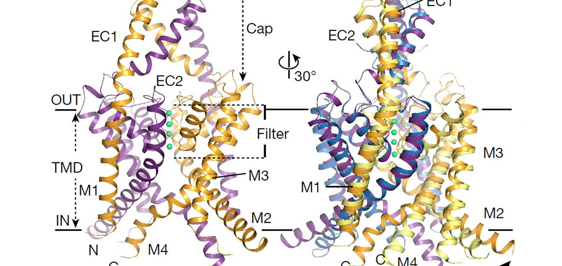
Living organisms perceive and respond to a diverse range of mechanical stimuli. A variety of mechanosensitive ion channels have evolved to facilitate these responses, but the molecular mechanisms underlying their exquisite sensitivity to different forces within the membrane remains unclear. TREK-2 is a mammalian two-pore domain (K2P) K+ channel important for mechanosensation, and recent studies have shown how increased membrane tension favors a more expanded conformation of the channel within the membrane. These channels respond to a complex range of mechanical stimuli, however, and it is uncertain how differences in tension between the inner and outer leaflets of the membrane contribute to this process. To examine this, we have combined computational approaches with functional studies of oppositely oriented single channels within the same lipid bilayer. Our results reveal how the asymmetric structure of TREK-2 allows it to distinguish a broad profile of forces within the membrane, and illustrate the mechanisms that eukaryotic mechanosensitive ion channels may use to detect and fine-tune their responses to different mechanical stimuli.The effects of stretch activation on ionic selectivity of the TREK-2 K2P K(+) channel
Channels Taylor and Francis 11:5 (2017) 482-486
Abstract:
The TREK-2 (KCNK10) K2P potassium channel can be regulated by variety of polymodal stimuli including pressure. In a recent study, we demonstrated that this mechanosensitive K(+) channel responds to changes in membrane tension by undergoing a major structural change from its 'down' state to the more expanded 'up' state conformation. These changes are mostly restricted to the lower part of the protein within the bilayer, but are allosterically coupled to the primary gating mechanism located within the selectivity filter. However, any such structural changes within the filter also have the potential to alter ionic selectivity and there are reports that some K2Ps, including TREK channels, exhibit a dynamic ionic selectivity. In this addendum to our previous study we have therefore examined whether the selectivity of TREK-2 is altered by stretch activation. Our results reveal that the filter remains stable and highly selective for K(+) over Na(+) during stretch activation, and that permeability to a range of other cations (Rb(+), Cs(+) and NH4(+)) also does not change. The asymmetric structural changes that occur during stretch activation therefore allow the channel to respond to changes in membrane tension without a loss of K(+) selectivity.Dynamic role of the tether helix in PIP2-dependent gating of a G protein-gated potassium channel
Journal of General Physiology Rockefeller University Press 149:8 (2017) 799-811
Abstract:
G protein-gated inwardly rectifying potassium (GIRK) channels control neuronal excitability in the brain and are implicated in several different neurological diseases. The anionic phospholipid phosphatidylinositol 4,5 bisphosphate (PIP2) is an essential cofactor for GIRK channel gating, but the precise mechanism by which PIP2 opens GIRK channels remains poorly understood. Previous structural studies have revealed several highly conserved, positively charged residues in the "tether helix" (C-linker) that interact with the negatively charged PIP2 However, these crystal structures of neuronal GIRK channels in complex with PIP2 provide only snapshots of PIP2's interaction with the channel and thus lack details about the gating transitions triggered by PIP2 binding. Here, our functional studies reveal that one of these conserved basic residues in GIRK2, Lys200 (6'K), supports a complex and dynamic interaction with PIP2 When Lys200 is mutated to an uncharged amino acid, it activates the channel by enhancing the interaction with PIP2 Atomistic molecular dynamic simulations of neuronal GIRK2 with the same 6' substitution reveal an open GIRK2 channel with PIP2 molecules adopting novel positions. This dynamic interaction with PIP2 may explain the intrinsic low open probability of GIRK channels and the mechanism underlying activation by G protein Gβγ subunits and ethanol.Bilayer-Mediated Structural Transitions Control Mechanosensitivity of the TREK-2 K2P Channel.
Structure Cell Press (2017)