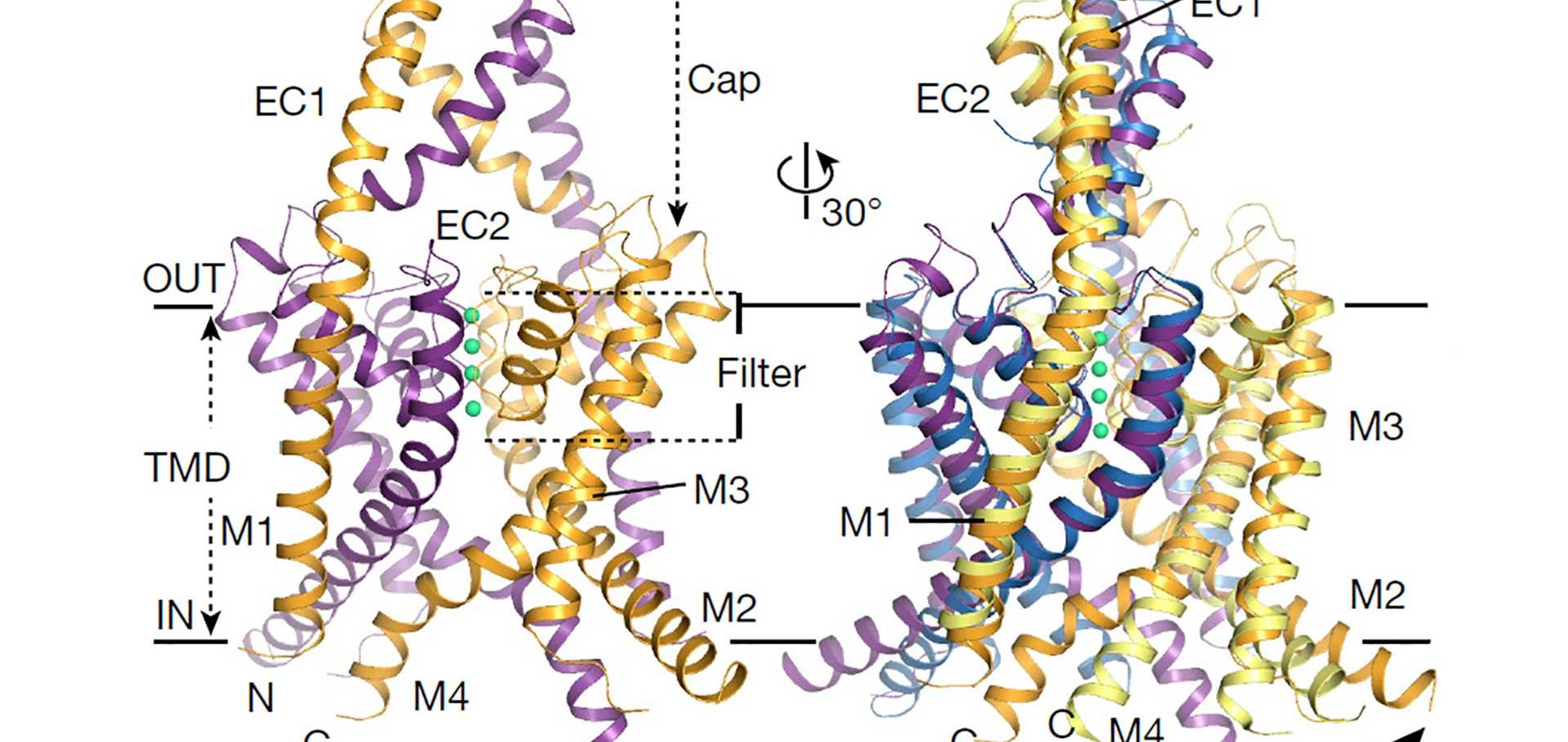
Structure of a KirBac potassium channel with an open bundle crossing indicates a mechanism of channel gating.
Nat Struct Mol Biol 19:2 (2012) 158-163
Abstract:
KirBac channels are prokaryotic homologs of mammalian inwardly rectifying (Kir) potassium channels, and recent crystal structures of both Kir and KirBac channels have provided major insight into their unique structural architecture. However, all of the available structures are closed at the helix bundle crossing, and therefore the structural mechanisms that control opening of their primary activation gate remain unknown. In this study, we engineered the inner pore-lining helix (TM2) of KirBac3.1 to trap the bundle crossing in an apparently open conformation and determined the crystal structure of this mutant channel to 3.05 Å resolution. Contrary to previous speculation, this new structure suggests a mechanistic model in which rotational 'twist' of the cytoplasmic domain is coupled to opening of the bundle-crossing gate through a network of inter- and intrasubunit interactions that involve the TM2 C-linker, slide helix, G-loop and the CD loop.Comparison of the Structure of a Bacterial Potassium Channel in Both 2D and 3D Crystals
BIOPHYSICAL JOURNAL 102:3 (2012) 536A-536A
Crystal Structure of a Prokaryotic Kir Channel in an Open Conformation
BIOPHYSICAL JOURNAL 102:3 (2012) 536A-536A
Functional analysis of missense variants in the TRESK (KCNK18) K channel.
Sci Rep 2 (2012) 237
Abstract:
A loss of function mutation in the TRESK K2P potassium channel (KCNK18), has recently been linked with typical familial migraine with aura. We now report the functional characterisation of additional TRESK channel missense variants identified in unrelated patients. Several variants either had no apparent functional effect, or they caused a reduction in channel activity. However, the C110R variant was found to cause a complete loss of TRESK function, yet is present in both sporadic migraine and control cohorts, and no variation in KCNK18 copy number was found. Thus despite the previously identified association between loss of TRESK channel activity and migraine in a large multigenerational pedigree, this finding indicates that a single non-functional TRESK variant is not alone sufficient to cause typical migraine and highlights the genetic complexity of this disorder.State-independent intracellular access of quaternary ammonium blockers to the pore of TREK-1.
Channels (Austin) 6:6 (2012) 473-478