Influence of lipids on the hydrophobic barrier within the pore of the TWIK-1 K2P channel
Channels Taylor and Francis 9:1 (2014) 44-49
Abstract:
Several recent ion channel structures have revealed large side portals, or ‘fenestrations’ at the interface between their transmembrane helices that potentially expose the ion conduction pathway to the lipid core of the bilayer. In a recent study we demonstrated that functional activity of the TWIK-1 K2P channel is influenced by the presence of hydrophobic residues deep within the inner pore. These residues are located near the fenestrations in the TWIK-1 structure and promote dewetting of the pore by forming a hydrophobic barrier to ion conduction. During our previous MD simulations, lipid tails were observed to enter these fenestrations. In this addendum to that study, we investigate lipid contribution to the dewetting process. Our results demonstrate that lipid tails from both the upper and lower leaflets occupy the fenestrations and penetrate into the pore. The lipid tails do not sterically occlude the pore, but there is an inverse correlation between the presence of water within the hydrophobic barrier and the number of lipids tails within the lining of the pore. However, dewetting still occurs in the absence of lipids tails, and pore hydration appears to be determined primarily by those side-chains lining the narrowest part of the pore cavity.Hydrophobic Gating in Ion Channels
Journal of Molecular Biology (2014)
Abstract:
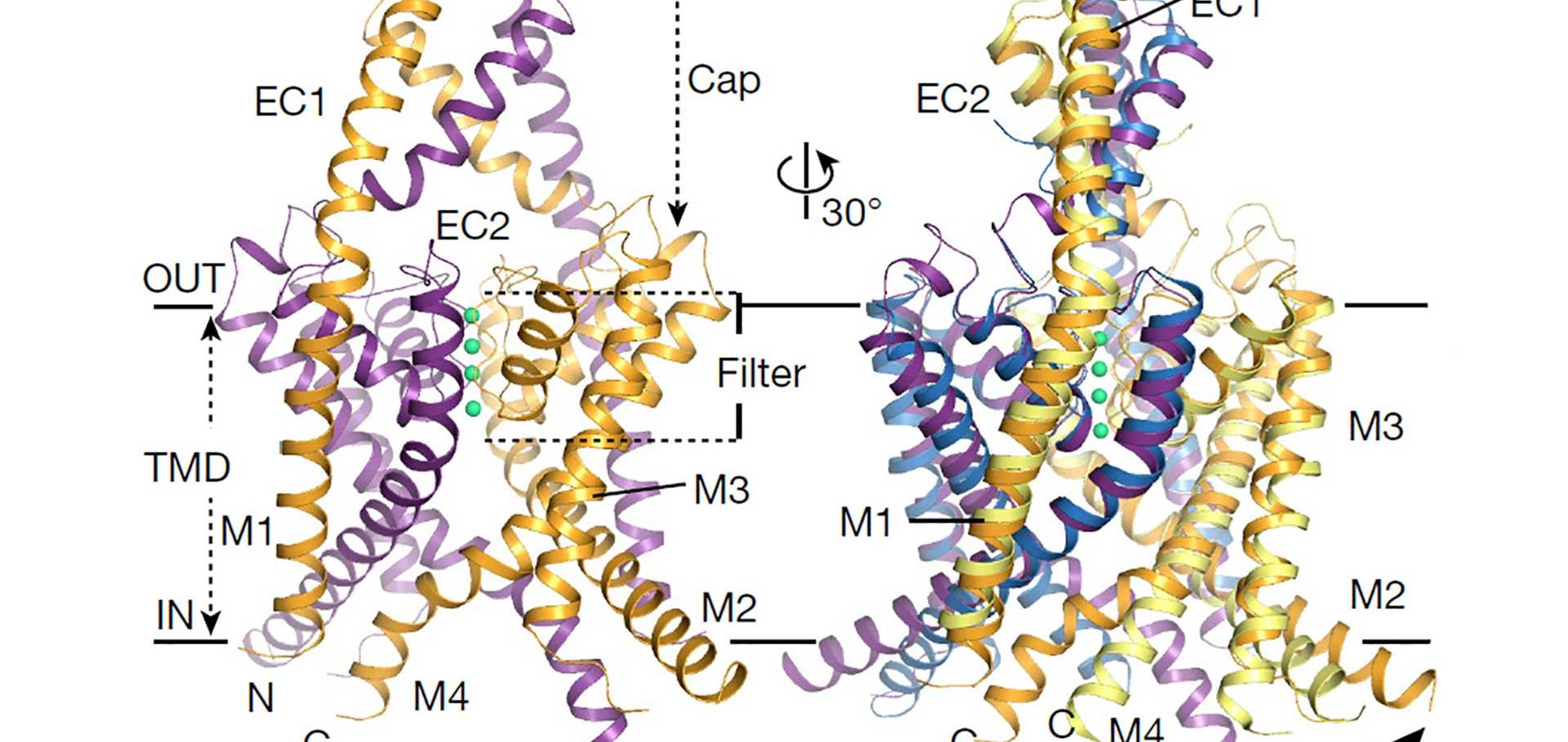
Biological ion channels are nanoscale transmembrane pores. When water and ions are enclosed within the narrow confines of a sub-nanometer hydrophobic pore, they exhibit behavior not evident from macroscopic descriptions. At this nanoscopic level, the unfavorable interaction between the lining of a hydrophobic pore and water may lead to stochastic liquid-vapor transitions. These transient vapor states are "dewetted", i.e. effectively devoid of water molecules within all or part of the pore, thus leading to an energetic barrier to ion conduction. This process, termed "hydrophobic gating", was first observed in molecular dynamics simulations of model nanopores, where the principles underlying hydrophobic gating (i.e., changes in diameter, polarity, or transmembrane voltage) have now been extensively validated. Computational, structural, and functional studies now indicate that biological ion channels may also exploit hydrophobic gating to regulate ion flow within their pores. Here we review the evidence for this process and propose that this unusual behavior of water represents an increasingly important element in understanding the relationship between ion channel structure and function. © 2014.Structures of the human two-pore domain potassium channels TREK-1 and TREK-2
Acta Crystallographica Section A: Foundations and advances International Union of Crystallography (IUCr) 70:a1 (2014) c1489-c1489
A hydrophobic barrier deep within the inner pore of the TWIK-1 K2P potassium channel
Nature Communications Springer Nature 5 (2014) 4377
Abstract:
Recent X-ray crystal structures of the two-pore domain (K2P) family of potassium channels have revealed a unique structural architecture at the point where the cytoplasmic bundle-crossing gate is found in most other tetrameric K+ channels. However, despite the apparently open nature of the inner pore in the TWIK-1 (K2P1/KCNK1) crystal structure, the reasons underlying its low levels of functional activity remain unclear. In this study, we use a combination of molecular dynamics simulations and functional validation to demonstrate that TWIK-1 possesses a hydrophobic barrier deep within the inner pore, and that stochastic dewetting of this hydrophobic constriction acts as a major barrier to ion conduction. These results not only provide an important insight into the mechanisms which control TWIK-1 channel activity, but also have important implications for our understanding of how ion permeation may be controlled in similar ion channels and pores.State-dependent network connectivity determines gating in a K+ channel
Structure 22:7 (2014) 1037-1046