DNA origami signposts for identifying proteins on cell membranes by electron cryotomography
Cell Cell Press 184:4 (2021) 1110-1121.e16
Abstract:
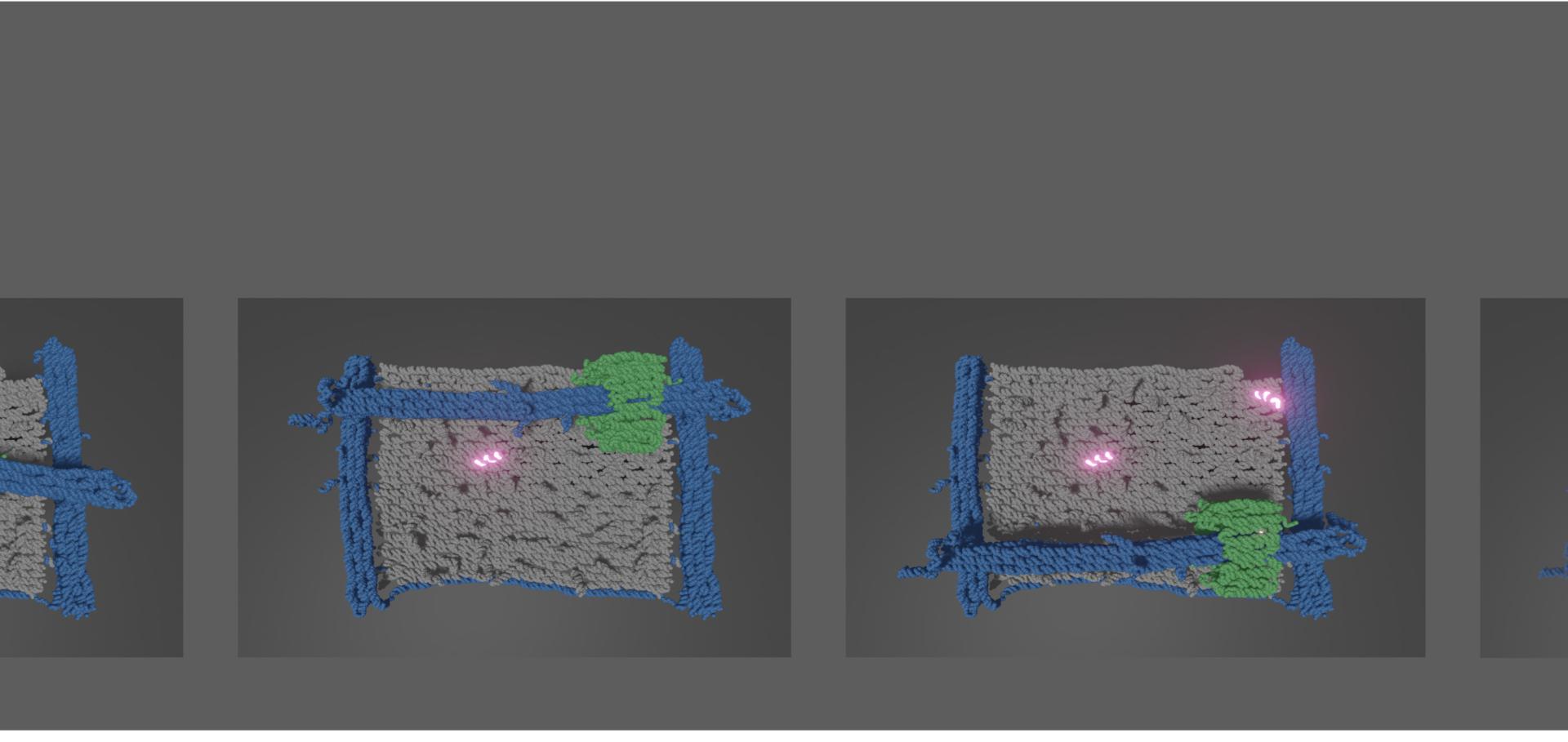
Electron cryotomography (cryoET), an electron cryomicroscopy (cryoEM) modality, has changed our understanding of biological function by revealing the native molecular details of membranes, viruses, and cells. However, identification of individual molecules within tomograms from cryoET is challenging because of sample crowding and low signal-to-noise ratios. Here, we present a tagging strategy for cryoET that precisely identifies individual protein complexes in tomograms without relying on metal clusters. Our method makes use of DNA origami to produce “molecular signposts” that target molecules of interest, here via fluorescent fusion proteins, providing a platform generally applicable to biological surfaces. We demonstrate the specificity of signpost origami tags (SPOTs) in vitro as well as their suitability for cryoET of membrane vesicles, enveloped viruses, and the exterior of intact mammalian cells.Reconfigurable T‐junction DNA origami
Angewandte Chemie International Edition Wiley 59:37 (2020) 15942-15946
Abstract:
DNA self‐assembly allows the construction of nanometre‐scale structures and devices. Structures with thousands of unique components are routinely assembled in good yield. Experimental progress has been rapid, based largely on empirical design rules. Here we demonstrate a DNA origami technique designed as a model system with which to explore the mechanism of assembly. The origami fold is controlled through single‐stranded loops embedded in a double‐stranded DNA template and is programmed by a set of double‐stranded linkers that specify pairwise interactions between loop sequences. Assembly is via T‐junctions formed by hybridization of single‐stranded overhangs on the linkers with the loops. The sequence of loops on the template and the set of interaction rules embodied in the linkers can be reconfigured with ease. We show that a set of just two interaction rules can be used to assemble simple T‐junction origami motifs and that assembly can be performed at room temperature.Reconfigurable T‐junction DNA origami
Angewandte Chemie International Edition Wiley (2020) anie.202006281
Reconfigurable T‐junction DNA origami
Angewandte Chemie Wiley (2020) ange.202006281
Characterising DNA T-motifs by Simulation and Experiment
(2020)