Design of hidden thermodynamic driving for non-equilibrium systems via mismatch elimination during DNA strand displacement
Abstract:
Recent years have seen great advances in the development of synthetic self-assembling molecular systems. Designing out-of-equilibrium architectures, however, requires a more subtle control over the thermodynamics and kinetics of reactions. We propose a mechanism for enhancing the thermodynamic drive of DNA strand-displacement reactions whilst barely perturbing forward reaction rates: the introduction of mismatches within the initial duplex. Through a combination of experiment and simulation, we demonstrate that displacement rates are strongly sensitive to mismatch location and can be tuned by rational design. By placing mismatches away from duplex ends, the thermodynamic drive for a strand-displacement reaction can be varied without significantly affecting the forward reaction rate. This hidden thermodynamic driving motif is ideal for the engineering of non-equilibrium systems that rely on catalytic control and must be robust to leak reactions.Controlling the bioreceptor spatial distribution at the nanoscale for single molecule counting in microwell arrays
Abstract:
The ability to detect low concentrations of protein biomarkers is crucial for the early-stage detection of many diseases and therefore indispensable for improving diagnostic devices for healthcare. Here, we demonstrate that by integrating DNA nanotechnologies like DNA origami and aptamers, we can design innovative biosensing concepts for reproducible and sensitive detection of specific targets. DNA origami structures decorated with aptamers were studied as a novel tool to structure the biosensor surface with nanoscale precision in a digital detection bioassay, enabling control of the density, orientation, and accessibility of the bioreceptor to optimize the interaction between target and aptamer. DNA origami was used to control the spatial distribution of an in-house-generated aptamer on superparamagnetic microparticles, resulting in an origami-linked digital aptamer bioassay to detect the main peanut antigen Ara h1 with 2-fold improved signal-to-noise ratio and 15-fold improved limit of detection compared to a digital bioassay without DNA origami. Moreover, the sensitivity achieved was 4 orders of magnitude higher than commercially available and literature-reported enzyme-linked immunosorbent assay techniques. In conclusion, this novel and innovative approach to engineer biosensing interfaces will be of major interest to scientists and clinicians looking for new molecular insights and ultrasensitive detection of a broad range of targets, and, for the next generation of diagnostics.Peptide assembly directed and quantified using megadalton DNA nanostructures
Abstract:
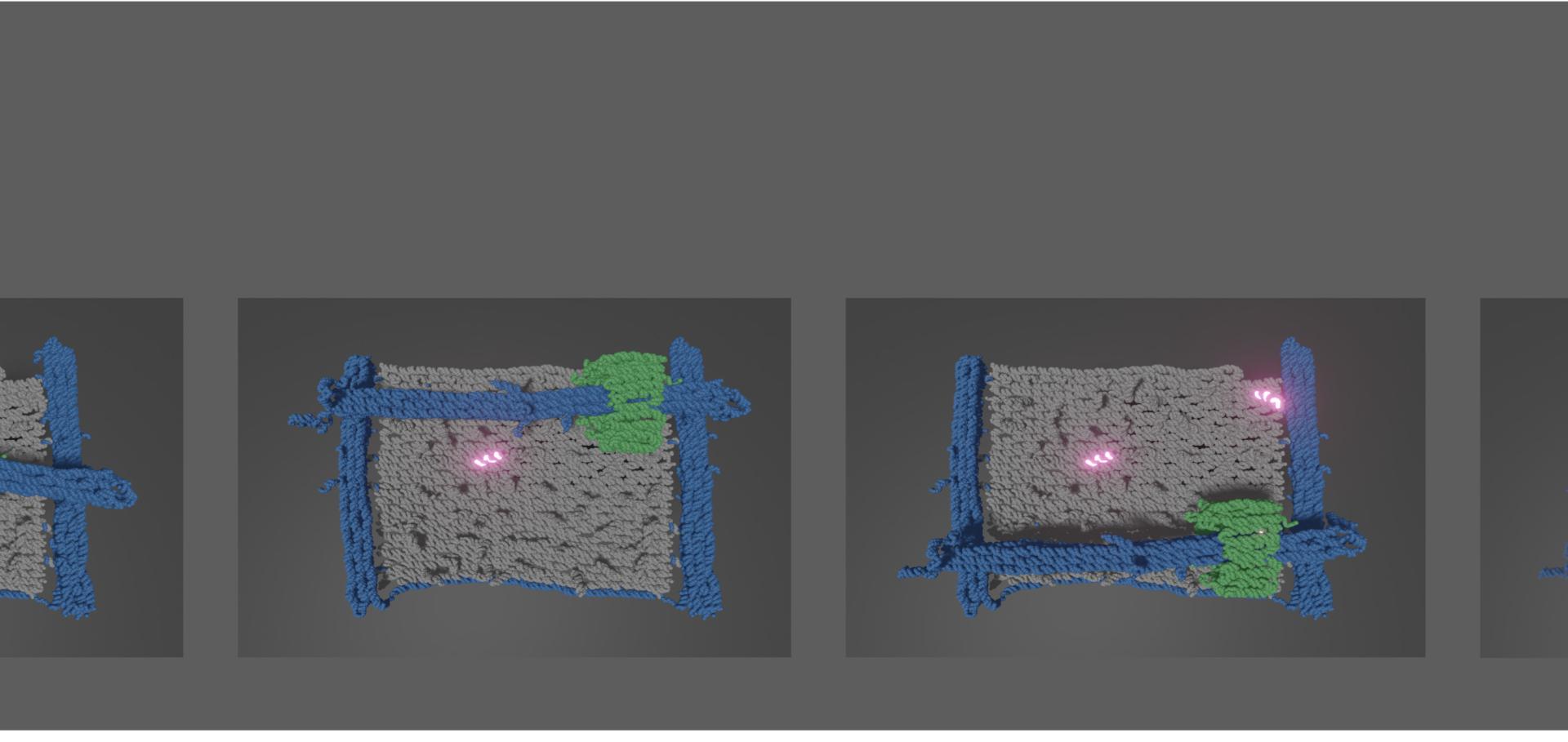
In nature, co-assembly of polypeptides, nucleic acids, and polysaccharides is used to create functional supramolecular structures. Here, we show that DNA nanostructures can be used to template interactions between peptides and to enable the quantification of multivalent interactions that would otherwise not be observable. Our functional building blocks are peptide–oligonucleotide conjugates comprising de novo designed dimeric coiled-coil peptides covalently linked to oligonucleotide tags. These conjugates are incorporated in megadalton DNA origami nanostructures and direct nanostructure association through peptide–peptide interactions. Free and bound nanostructures can be counted directly from electron micrographs, allowing estimation of the dissociation constants of the peptides linking them. Results for a single peptide–peptide interaction are consistent with the measured solution-phase free energy; DNA nanostructures displaying multiple peptides allow the effects of polyvalency to be probed. This use of DNA nanostructures as identifiers allows the binding strengths of homo- and heterodimeric peptide combinations to be measured in a single experiment and gives access to dissociation constants that are too low to be quantified by conventional techniques. The work also demonstrates that hybrid biomolecules can be programmed to achieve spatial organization of complex synthetic biomolecular assemblies.