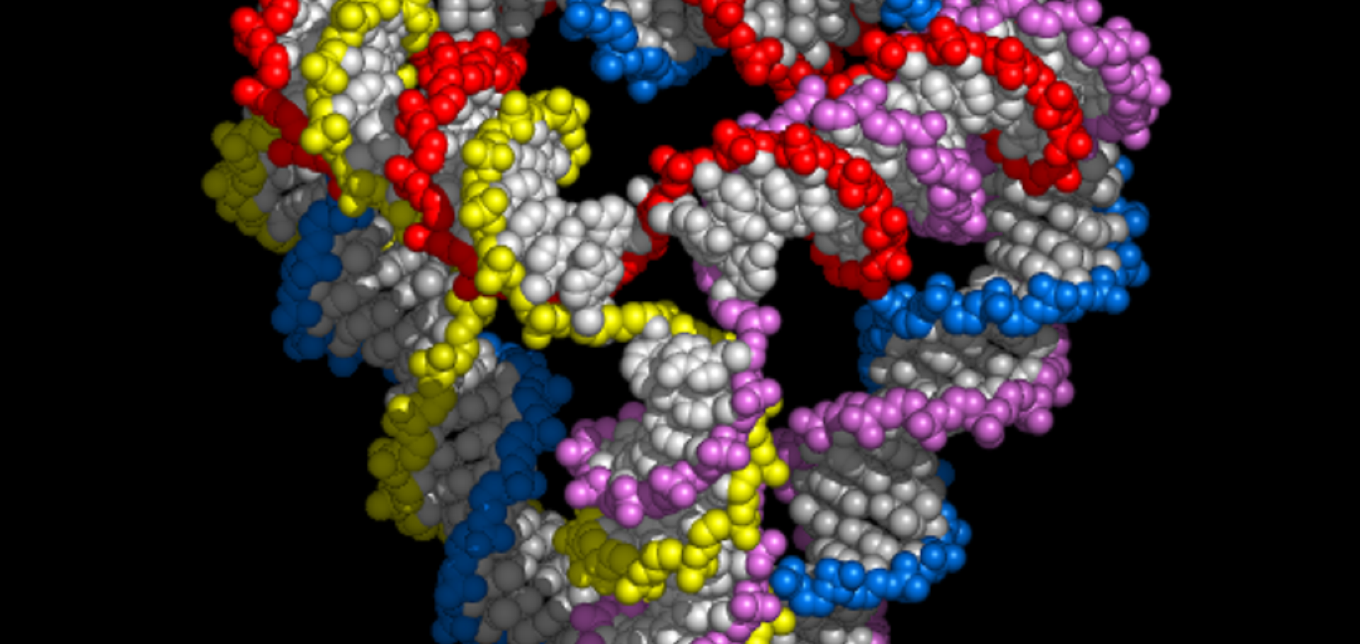
Home
Department Of Physics text logo
  • Research
    • Our research
    • Our research groups
    • Our research in action
    • Research funding support
    • Summer internships for undergraduates
  • Study
    • Undergraduates
    • Postgraduates
  • Engage
    • For alumni
    • For business
    • For schools
    • For the public
Menu
DNA tetrahedron

Professor Andrew Turberfield

Professor of Biological Physics

Research theme

  • Biological physics

Sub department

  • Condensed Matter Physics

Research groups

  • Nucleic acid nanotechnology
Andrew.Turberfield@physics.ox.ac.uk
  • About
  • Publications

DNA-based optical sensors for forces in cytoskeletal networks

ACS Applied Nano Materials American Chemical Society 6:17 (2023) 15455-15464

Authors:

Christina Jayachandran, Arindam Ghosh, Meenakshi Prabhune, Jonathan Bath, Andrew JJ Turberfield, Lara Hauke, Jorg Enderlein, Florian Rehfeldt, Christoph FF Schmidt

Abstract:

Mechanical forces are relevant for many biological processes, from wound healing and tumor formation to cell migration and differentiation. Cytoskeletal actin is largely responsible for responding to forces and transmitting them in cells, while also maintaining cell shape and integrity. Here, we describe a FRET-based hybrid DNA-protein tension sensor that is designed to sample transient forces in actin networks by employing two actin-binding motifs with a fast off-rate attached to a central DNA hairpin loop. Such a sensor will be useful to monitor rapidly changing stresses in the cell cytoskeleton. We use fluorescence lifetime imaging to determine the FRET efficiency and thereby the conformational state of the sensor, which makes the measurement robust against intensity variations. We demonstrate the applicability of the sensor by confocal microscopy and by monitoring crosslinking activity in in vitro actin networks by bulk rheology.
More details from the publisher
Details from ORA
More details

A modular RNA delivery system comprising spherical nucleic acids built on endosome-escaping polymeric nanoparticles

Nanoscale Advances Royal Society of Chemistry 5 (2023) 2941-2949

Authors:

Antonio Garcia-Guerra, Ruth Ellerington, Jens Gaitzsch, Jonathan Bath, Mahnseok Kye, Miguel A Varela, Giuseppe Battaglia, Matthew JA Wood, Raquel Manzano, Carlo Rinaldi, Andrew J Turberfield

Abstract:

Nucleic acid therapeutics require delivery systems to reach their targets. Key challenges to be overcome include avoidance of accumulation in cells of the mononuclear phagocyte system and escape from the endosomal pathway. Spherical nucleic acids (SNAs), in which a gold nanoparticle supports a corona of oligonucleotides, are promising carriers for nucleic acids with valuable properties including nuclease resistance, sequence-specific loading and control of receptor-mediated endocytosis. However, SNAs accumulate in the endosomal pathway and are thus vulnerable to lysosomal degradation or recycling exocytosis. Here, an alternative SNA core based on diblock copolymer PMPC25–PDPA72 is investigated. This pH-sensitive polymer self-assembles into vesicles with an intrinsic ability to escape endosomes via osmotic shock triggered by acidification-induced disassembly. DNA oligos conjugated to PMPC25–PDPA72 molecules form vesicles, or polymersomes, with DNA coronae on luminal and external surfaces. Nucleic acid cargoes or nucleic acid-tagged targeting moieties can be attached by hybridization to the coronal DNA. These polymeric SNAs are used to deliver siRNA duplexes against C9orf72, a genetic target with therapeutic potential for amyotrophic lateral sclerosis, to motor neuron-like cells. By attaching a neuron-specific targeting peptide to the PSNA corona, effective knock-down is achieved at doses of 2 particles per cell.

More details from the publisher
Details from ORA
More details
More details

Designing the self-assembly of arbitrary shapes using minimal complexity building blocks

ACS Nano American Chemical Society 17:6 (2023) 5387-5398

Authors:

Joakim Bohlin, Andrew J Turberfield, Ard A Louis, Petr Šulc

Abstract:

The design space for self-assembled multicomponent objects ranges from a solution in which every building block is unique to one with the minimum number of distinct building blocks that unambiguously define the target structure. We develop a pipeline to explore the design spaces for a set of structures of various sizes and complexities. To understand the implications of the different solutions, we analyze their assembly dynamics using patchy particle simulations and study the influence of the number of distinct building blocks, and the angular and spatial tolerances on their interactions, on the kinetics and yield of the target assembly. We show that the resource-saving solution with a minimum number of distinct blocks can often assemble just as well (or faster) than designs where each building block is unique. We further use our methods to design multifarious structures, where building blocks are shared between different target structures. Finally, we use coarse-grained DNA simulations to investigate the realization of multicomponent shapes using DNA nanostructures as building blocks.
More details from the publisher
Details from ORA
More details
More details

Far-field electrostatic signatures of macromolecular 3D conformation

Nano Letters American Chemical Society 22:19 (2022) 7834-7840

Authors:

Gunnar Kloes, Timothy JD Bennett, Alma Chapet-Batlle, Ali Behjatian, Andrew Turberfield, Madhavi Krishnan

Abstract:

In solution as in vacuum, the electrostatic field distribution in the vicinity of a charged object carries information on its three-dimensional geometry. We report on an experimental study exploring the effect of molecular shape on long-range electrostatic interactions in solution. Working with DNA nanostructures carrying approximately equal amounts of total charge but each in a different three-dimensional conformation, we demonstrate that the geometry of the distribution of charge in a molecule has substantial impact on its electrical interactions. For instance, a tetrahedral structure, which is the most compact distribution of charge we tested, can create a far-field effect that is effectively identical to that of a rod-shaped molecule carrying half the amount of total structural charge. Our experiments demonstrate that escape-time electrometry (ETe) furnishes a rapid and facile method to screen and identify 3D conformations of charged biomolecules or molecular complexes in solution.
More details from the publisher
Details from ORA
More details
More details

A DNA molecular printer capable of programmable positioning and patterning in two dimensions

Science Robotics American Association for the Advancement of Science 7:65 (2022) eabn5459

Authors:

Erik Benson, rafael Carrascosa Marzo, jonathan Bath, Andrew Turberfield

Abstract:

Nanoscale manipulation and patterning usually require costly and sensitive top-down techniques such as those used in scanning probe microscopies or in semiconductor lithography. DNA nanotechnology enables exploration of bottom-up fabrication and has previously been used to design self-assembling components capable of linear and rotary motion. In this work, we combine three independently controllable DNA origami linear actuators to create a nanoscale robotic printer. The two-axis positioning mechanism comprises a moveable gantry, running on parallel rails, threading a mobile sleeve. We show that the device is capable of reversibly positioning a write head over a canvas through the addition of signaling oligonucleotides. We demonstrate “write” functionality by using the head to catalyze a local DNA strand–exchange reaction, selectively modifying pixels on a canvas. This work demonstrates the power of DNA nanotechnology for creating nanoscale robotic components and could find application in surface manufacturing, biophysical studies, and templated chemistry.
More details from the publisher
Details from ORA
More details
More details

Pagination

  • First page First
  • Previous page Prev
  • Page 1
  • Page 2
  • Current page 3
  • Page 4
  • Page 5
  • Page 6
  • Page 7
  • Page 8
  • Page 9
  • …
  • Next page Next
  • Last page Last

Footer Menu

  • Contact us
  • Giving to the Dept of Physics
  • Work with us
  • Media

User account menu

  • Log in

Follow us

FIND US

Clarendon Laboratory,

Parks Road,

Oxford,

OX1 3PU

CONTACT US

Tel: +44(0)1865272200

University of Oxfrod logo Department Of Physics text logo
IOP Juno Champion logo Athena Swan Silver Award logo

© University of Oxford - Department of Physics

Cookies | Privacy policy | Accessibility statement

Built by: Versantus

  • Home
  • Research
  • Study
  • Engage
  • Our people
  • News & Comment
  • Events
  • Our facilities & services
  • About us
  • Current students
  • Staff intranet